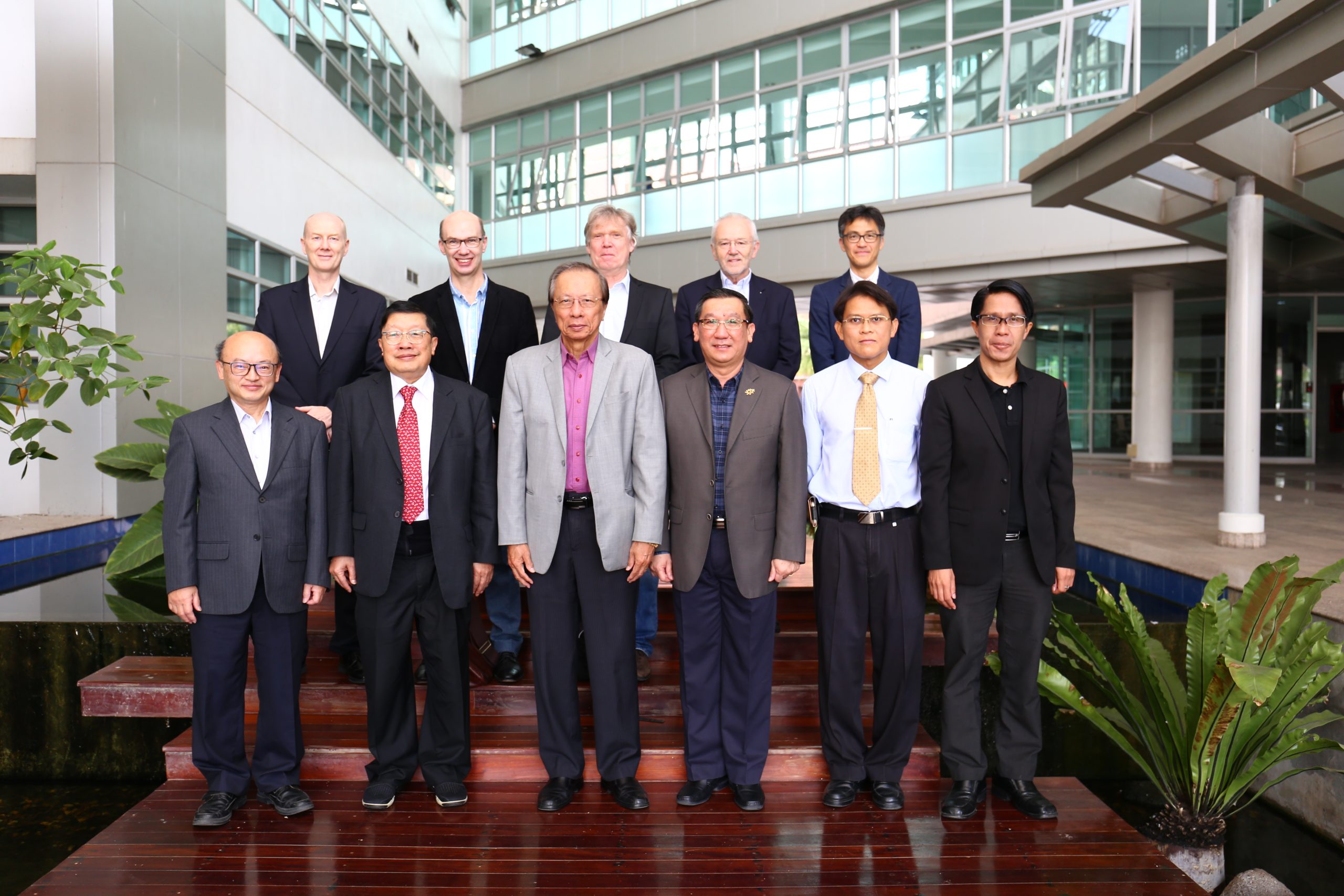
Professor Wing Commander Dr. Sarawut Sujitjorn, Chief Executive Officer of Synchrotron Light Research Institute (SLRI), and executives welcomed SLRI International Advisory Committee (IAC) coming for a meeting on 18-19 March 2019 at SLRI.
The IAC came for the meeting to follow up work progress at SLRI in terms of accelerator operation, beamline implementation, user service provision, and provide SLRI with constructive comments, suggestions, and ideas to improve operation and service. The IAC is the committee which has played an important role in supporting implementation and work planning of SLRI.
Results of the meeting will be very supportive and useful for SLRI to develop related operation, implementation and service to be the most efficient and effective for SLRI users.
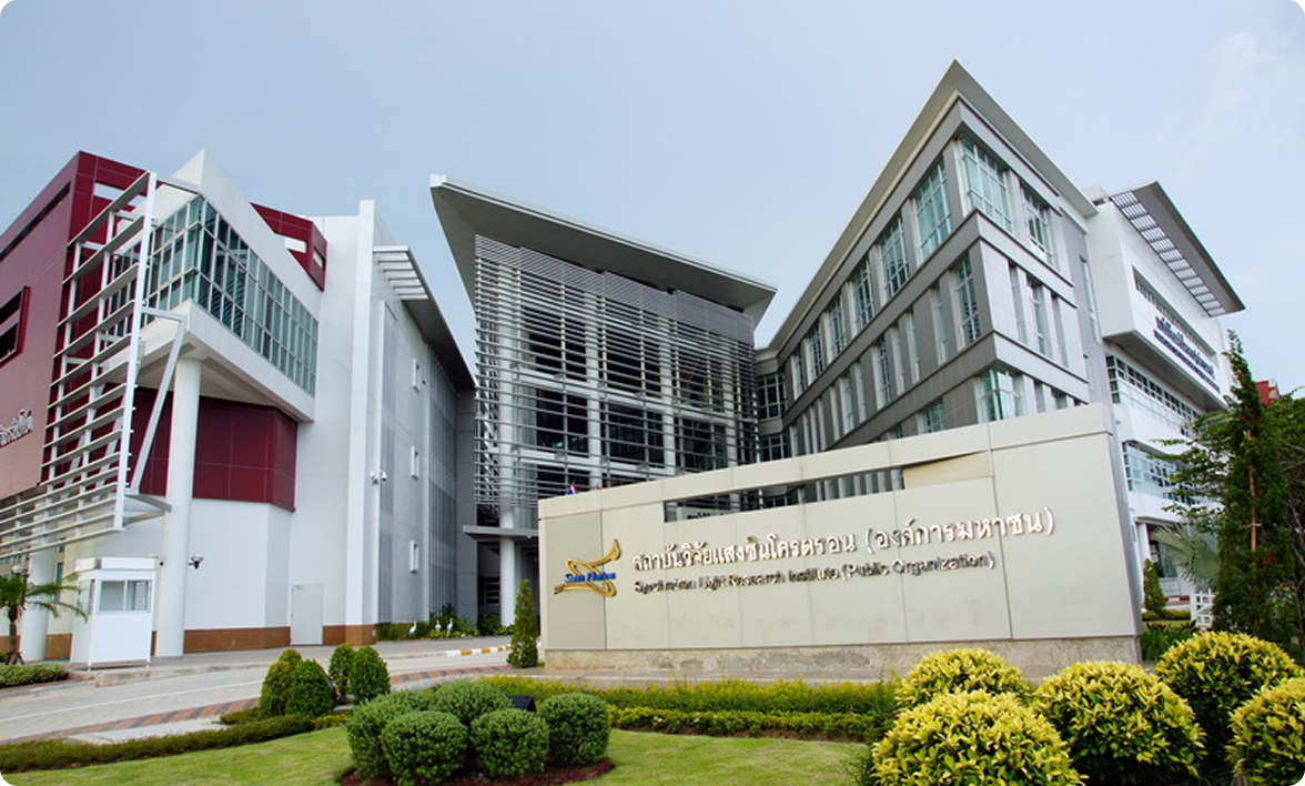
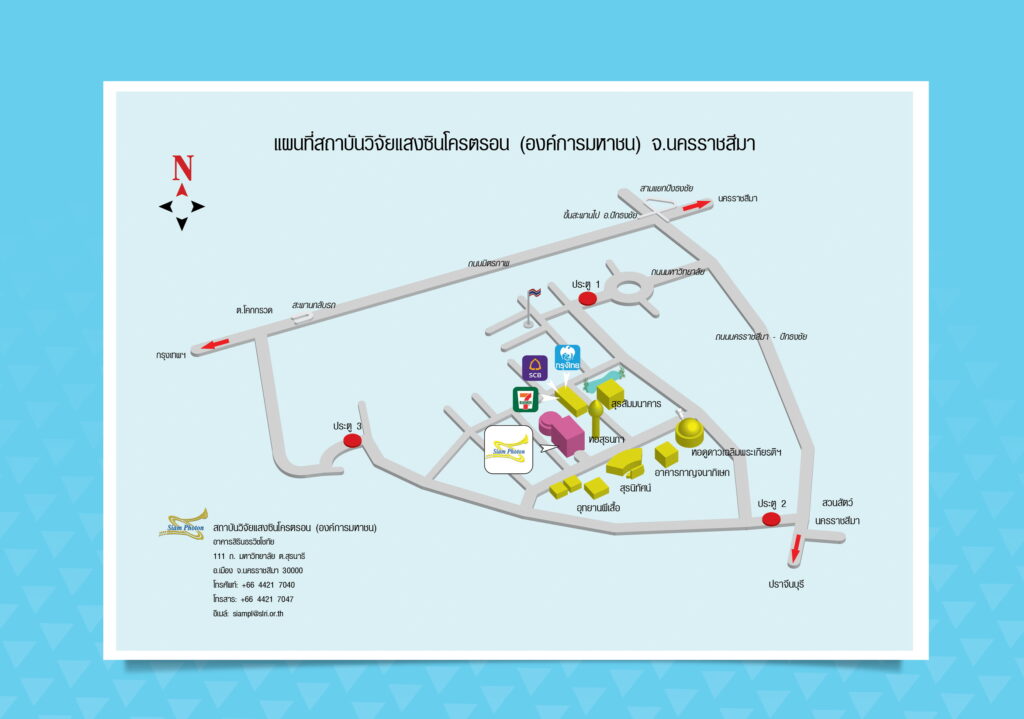
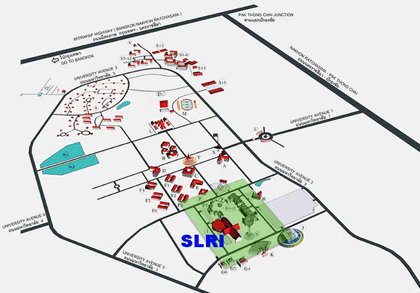
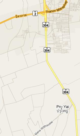
Synchrotron light research Institute located within the suranaree University of technology, which is located along the Nakhon Ratchasima – Pak Thong Chai road (line 304)
Show location synchrotron light research Institute within the area, suranaree University of technology
Travel by car,
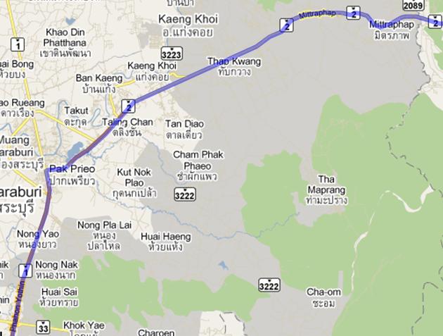
directions from Bangkok to Nakhon Ratchasima, there are 2 main path is the path phaholyothin – friendship and route Bangkok – Chachoengsao – Nakhon Ratchasima
route phaholyothin – friendship
, you can use Vibhavadi-Rangsit road, paholyothin road, or use ring road, Eastern or Western ring paholyothin road, at different levels, Bang PA-in to aim to Saraburi and then Saraburi turn right onto friendship road towards Nakhon Ratchasima
About 17 km before reaching the town of Nakhon Ratchasima to a separate level, Nakhon Ratchasima-Khon Kaen-Nakhon Ratchasima left out into the interchange, turn right towards Pak Thong Chai road (line 304), then about 7 km from the junction turn right onto University Avenue, another 5 kilometers to the suranaree University of technology, the distance from Bangkok to Nakhon Ratchasima 250 kilometers and takes about 3 hours.
The route Bangkok – Chachoengsao – Nakhon Ratchasima
from suwinthawong road towards Chachoengsao (road 304) along the road 304 through Chachoengsao to Nakhon Ratchasima via Pak Thong Chai to about 20 km, it will reach a junction, turn left onto University Avenue, towards the suranaree University of technology for this route takes about 3 hours and 45 minutes
travel by bus
You can take a bus from mo chit bus terminal (new bus station North-northeast) geared towards Nakhon Ratchasima bus terminal (bus terminal. New) bus. 1 journey to the. Nakhon Ratchasima 3, the company is air Korat development Ratchasima tour and suranaree air by leaving every half hour, 24 hours bus trip takes approximately 3 hours and 30 minutes, fare 198 million from Nakhon Ratchasima bus terminal travel synchrotron light research Institute, which is located in the suranaree University of technology by bus(minibus) line 17 (color: white, purple), which will be parked at the transport suranaree University of technology (the fare is about 15 baht) or can travel by taxi (Fares, Mao 250 baht). taxi 0-4437-0999
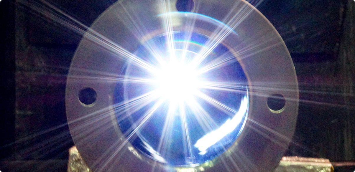
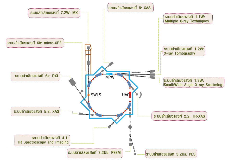
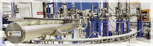
The storage ring is to store the electron beam for long period of time, and synchrotron radiation is emitted when electrons are curved in the magnetic field of the storage ring.
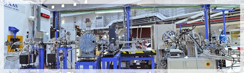
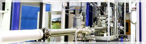
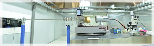
The synchrotron light (Beamline) is a group of devices that deliver energy electron from the band the retention of the electron to the experiment station at the synchrotron light generally consists of vacuum tube (vacuum tube) glass integrated light (collimating mirror) system selected energy light (stray) mirrors to focus light (focusing mirror) system (slit system) and other devices for customizable optical characteristics to requirements at the tip of the synchrotron light with experiment station, which has a system of measurement for experimental techniques specific
Currently, the Siam photon laboratory system, conveyor light 10 system for which has installed the experiment station. the 12 stations , both open, then, is that during the test, use the following:
1. The synchrotron light at 1.1 W: Multiple X-ray Texhniques
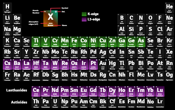
The synchrotron light at 1.1 W: Multiple X-ray Techniques (MXT) technique, absorption of x-rays (X-ray Absorption Spectroscopy, XAS) energy from 4 to 18 keV system conveyor light that combines experimental techniques that use x-rays for both absorption interference and fluorescent x-rays can provide information on the elemental composition, crystal structure and structure-level atoms of the sample.
2. The synchrotron light 1.2 W: X-ray Tomography
The synchrotron light 1.2 W: X-ray Imaging and X-ray Tomographic Microscopy (XTM) synchrotron light for CT imaging small objects is a system conveyor light can be if an image inside of an object, for example by using x-rays, and not destroy the sample can be analyzed aware of the composition of trace elements in the various conveyor system
3. The synchrotron light at 1.3 W: Small/Wide Angle X-ray Scattering
The synchrotron light at 1.3 W: Small/Wide Angle X-ray Scattering (SAXS/WAXS) for the study of the structure of nanometer scale by the technique of Small Angle X-ray Scattering system, conveyor light using the scattering of x-rays indicate the arrangement of the molecular size, shape, structure of the sample in nanometer
4. The synchrotron light 2.2: TR-XAS
The synchrotron light 2.2: Time-resolved X-ray Absorption Spectroscopy (TRXAS) for the study of the structure at an atomic level, by the technique of X-ray Absorption Spectroscopy system, conveyor optical techniques absorption of x-rays used to study the structure of the sample at the atomic level. following the change of the sample under the influence of environmental conditions such as temperature, pressure, gas, electric current and potential difference, because used in the study to develop chemical catalysts, fuel cells, batteries, etc.
5. The synchrotron light at 3.2 Ua/b: PES/PEEM
5.1 experimental station BL3. 2Ua: PES
Experiment station BL3. 2Ua: PES (Photoelectron Emission Spectroscopy) for the study of the electronic structure on the surface by the technique of Photoelectron Emission Spectroscopy is a technique for measuring the liberation of electrons by light, synchrotron tron in the near ultraviolet and x-rays, low energy, to study the energy value, the electrons come off the surface of the sample when the light synchrotron tron come incidence, such as the detection of adulteration and chemical structure, the surface area of the sample, etc.
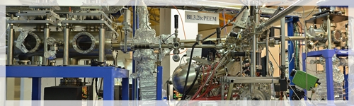
5.2 experimental station BL3. 2Ub: PEEM
Experiment station BL3. 2Ub: PEEM (Photoelectron Emission Microscopy) for imaging surface by the technique of Photoemission Electron Microscopy is a system conveyor light that can create an image from an electron release from the surface samples when the light synchrotron tron incidence may choose to take only the area you are interested in nanometer level, both also studied the chemical composition in the various areas that are shooting immediately, such as education, stripes and blame it on the metal surface corrosion of metal surfaces, various thin film and coating
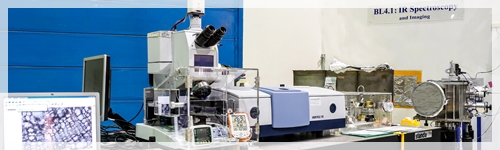
6. The synchrotron light 4.1: IR Spectroscopy and Imaging
The synchrotron light 4.1: Infrared Spectroscopy and Imaging (ISI) for the review and study the molecular structure of the substance by virtue of optical absorption in the near-infrared radiation is a technique optical absorption in the near infrared rays to analysis, monitoring and study of the structure among the atoms that show specific properties (functional group) of the molecule organic compound can classify the type of structure and the chemical bonds such as biomolecule polymer for the development of rubber products, food, medicine and cosmetics, can also be used microscopy techniques infrared in the study sample is small, such as cells, plant cells, animal hair and fiber, etc.
7. The synchrotron light at 5.2: XAS
The synchrotron light at 5.2: X-ray Absorption Spectroscopy (XAS), SUT-NANOTEC-SLRI XAS Beamline for the study of the structure at an atomic level, by the technique of X-ray Absorption Spectroscopy is a technique for absorption of x-rays to study the type and chemical composition of the elements present in the sample can also be told to arrange the booking atoms around, suitable for a variety of research, research, sample, either solid, liquid, and gas
8. The synchrotron light 6a/b: DXL/Micro-XRF
8.1 experimental station BL6a: DXL
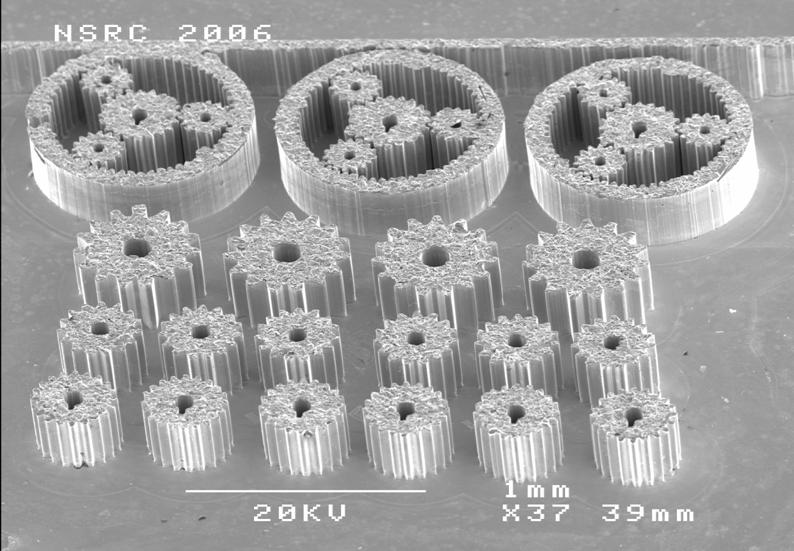
Experiment station BL6a: DXL (Deep X-ray Lithography) , for production parts, mechanical miniature size by the technique of Deep X-ray Lithography is a technique for bath x-rays, for production parts, comma, three-dimensional level µm (1 in 1,000 mm), this technique can be applied in various industries such as parts mechanical small gear clock sensor parts mould, comma, including the study of political change image when the object is illuminated with x-rays.
8.2 experimental station BL6b: micro-XRF
Experiment station BL6b: Micro-XRF (Micro-X-ray Fluorescence) for the study type and the distribution of elemental composition in the samples by the technique of micro-X-ray Fluorescence is a technique for bearing x-rays to monitor and track the composition of trace elements in the atomic level, such as stone, metal, or examples of creatures such as leaves, trees, etc., can also study the distribution of various trace elements, by creating a glowing x-rays
9. The synchrotron light 7.2 W: MX
The synchrotron light 7.2 W: Macromolecular Crystallography (MX) for the study of the structure of large molecules by the technique of Macromolecular Crystallography is a technique to study the three-dimensional structure of a biomolecule such various proteins can understand the structures and functions the function of the protein and various substances entering the reaction with the protein, which is of paramount importance to research in the fields of medicine, biology, such as improving the function of various enzymes, etc.
10. The synchrotron light at 8: XAS
The synchrotron light at 8: X-ray Absorption Spectroscopy (XAS) for the study of the structure at an atomic level, by the technique of X-ray Absorption Spectroscopy is a technique for absorption of x-rays for the study, the type of the elements and the arrangement of the atoms around the atom of the element of interest is also used to identify the oxidation state of the element, including study characteristics, chemical bonding, symmetry, area di Taser, etc. examples of research that can be done at the synchrotron light of this, such as ceramics, electric, additives, catalyst semiconductor, fuel cell, and Antiques, including trace elements, calcium, sulphur in natural product and phosphorus in the soil, etc.
For details, synchrotron light and experiment station, including contact Manager, synchrotron light, see in the page web of each of the synchrotron light
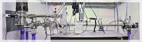
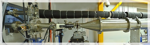
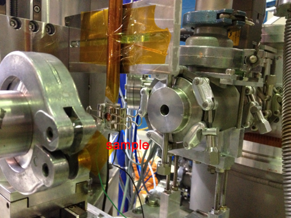
The primary advantage of the TRXAS end-station is its ability to record complete X-ray absorption spectra simultaneously and rapidly. This makes it an ideal tool for in-situ measurements to monitor structural and electronic changes in samples under various environmental conditions, such as high temperature, pressure, or specific gas atmospheres.
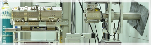
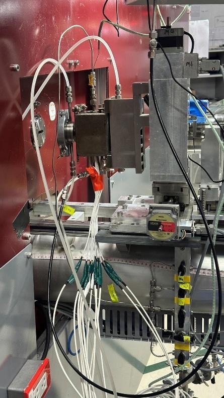
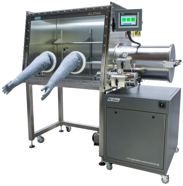
In-situ Environmental System
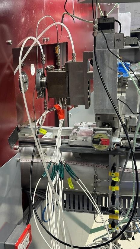
Currently, the institute provides a specialized Heating Cell designed to support a wide range of in-situ research applications, enabling simultaneous temperature control and atmospheric regulation.
Technical Specifications
Feature
Details
Material
Stainless Steel Body
Body Dimensions
45 x 45 x 70 mm
Sample Holder
16 mm diameter (pre-drilled hole)
Windows
Dual Kapton windows with water cooling system
Heating Source
6X Cartridge Heaters (8 mm OD, 250 W each)
Temperature Control
Maximum Operating Temperature
973 K (700 °C)
Ramp Rate
5-20 °C/min
Controller
Eurotherm 2216e with K-type thermocouple
Stability
± 0.1 K
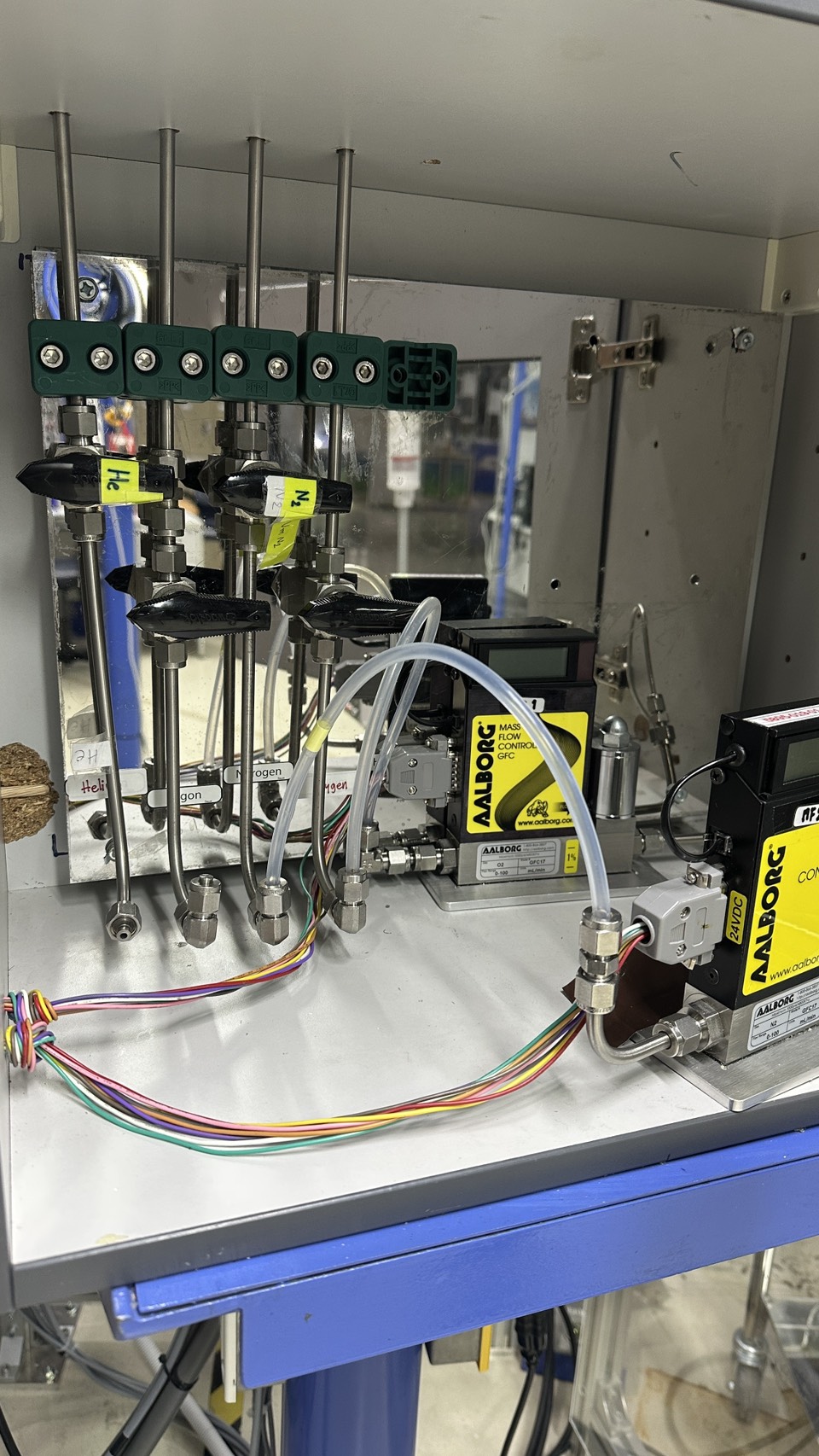
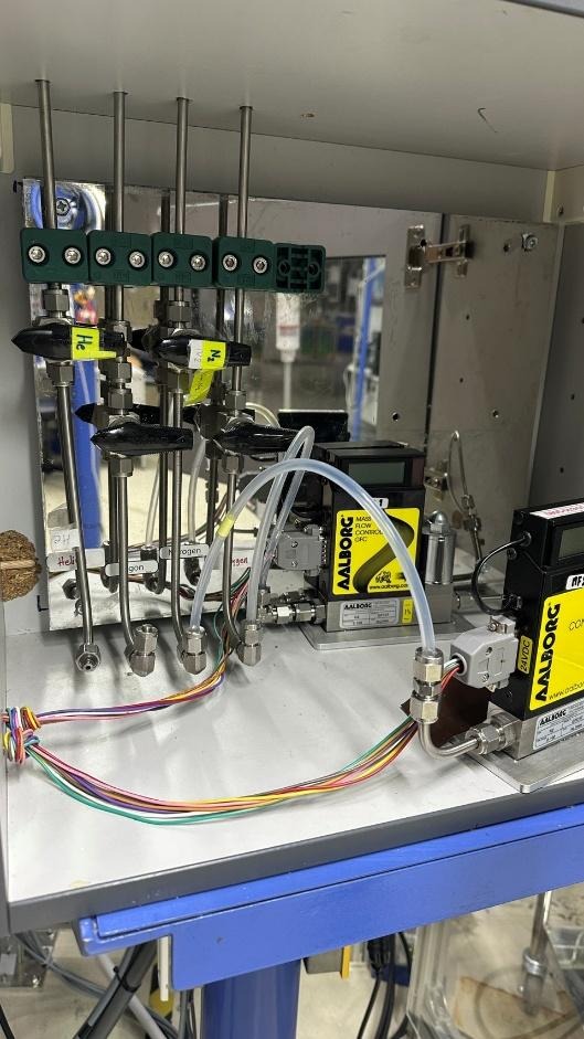
Gas Atmosphere & Flow Control
The cell is designed to accommodate various gas environments for catalytic and material study.
Available Gases
N2, O2, He, Ar, CO2 and H2
Flow Control
AALBORG Mass Flow Controllers (MFCs)
Flow Rate Ranges
0–100 sccm and 0–25 sccm
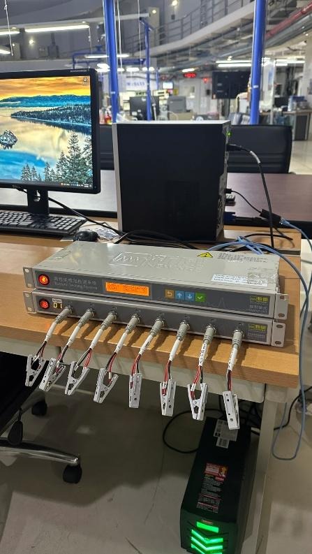
In-situ Battery System
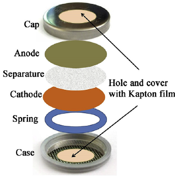
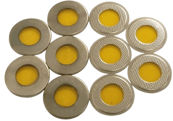
The cell structure consisted of a cap, anode, separator, cathode, spring, and case. The coin cell (CR2032) with an OD of 20 mm was used. To assembling the cell, an anode (Li foil, 0.75 mm thickness), separator (Celgard2400, 25 μm thickness), cathode (LFP) and spring (stainless steel, 0.3 mm thickness) were provided. Since the case and cap were made from stainless steel, they did not allow x-ray transmission. Thus, the aperture with OD 8 mm and sealed with Kapton film were provided.
Coin Cell Analyzer 5V10mA Battery Testing System
Technical Specifications
AC Input
AC 220V±10% 50Hz
Power
25 W
Resolution
AD: 16bit; DA: 12bit
Input
≥1MΩ
Voltage
Measuring Range
Charge:25mV~5V
Discharge: 25mV~5V
Discharge Min Voltage
0V
Accuracy
± 0.1% of FS
Stability
± 0.1% of FS
Current
Range
Charge: 0.05mA~10mA
Discharge: 0.05mA~10mA
Accuracy
± 0.1% of FS
Stability
± 0.1% of FS
Power
Output Power Per Channel
0.05W
Stability
± 0.2% of FS
Time
Rise Time
1ms (0~Full Range)
Step Time
≤ (365*24) hour/step
Time Format 00:00:00 (h:min:s)
Data Acquisition
Intervals
Time interval Δt:(1s~60000s)
Voltage interval ΔU: (10mV~5V)
Current interval ΔI: (0.02mA~10mA)
Frequency
1Hz
Charge
Mode of Operation
CCC, CVC, CC & CVC
End Conditions
Voltage, Current, Test Time, Capacity
Discharge
Mode of Operation
CCD, CPD
End Conditions
Voltage, Current, Test Time, Capacity
Cycle
Cycles
65535
Steps
63
Nested Function
Max three levels of loops
Detector System
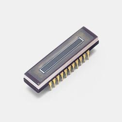
To measure XAS spectrum, the position sensitive detector (PSD) is used.
S3904–1024F
NMOS Linear Image Sensor with Scintillator
(Hamamatsu Photonics K.K.)
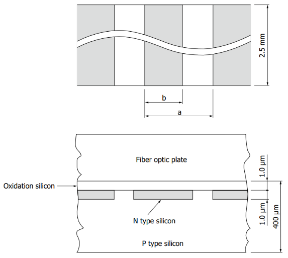
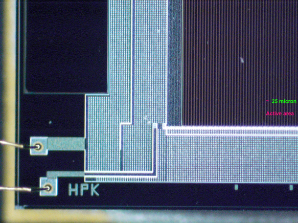
NMOS linear image sensor consisting of 1024 pixels of an N-type diffusion layer formed on a P-type silicon substrate.
Detection Method
The use of an indirect detection method, employing an X-ray scintillator (150 μm thick CsI (Cesium Iodide) coated on a fiber optic plate) to convert X-rays into visible light, extended the sensor lifetime to over a year.
This page provides an overview of the steps involved in preparing, submitting, and carrying out an experiment on BL2.2. Please explore the sub-sections for detailed guidance. If you have any questions or require additional information, you are encouraged to contact the BL8 beamline staff for assistance.
Access routes
Standard Access
Synchrotron Thailand runs three main Calls for Proposals each year. Proposals submitted via this route are evaluated by an independent peer review panel and successful applicants are allocated beamtime in the regular schedule. We strongly recommend contacting BL8 beamline staff before submission to discuss feasibility and the most suitable experimental configuration.
How to apply: submit your proposal through the official user application system and follow the deadlines listed on the Synchrotron Thailand website. For pre-submission advice, contact the Proposals Office.
Rapid Access
Rapid Access is intended for short, urgent, or time-sensitive ex-situ experiments for example, proof of concept measurements or quick data required for responding to referees. Rapid Access requests are considered outside the main Calls and are assessed on a case by case basis.
How to apply / who to contact: contact the Head, X-ray Spectroscopy section or beamline staff prior to submitting a Rapid Access request so the team can advise on feasibility and prioritisation. All Rapid Access applications must be submitted through the user application system.
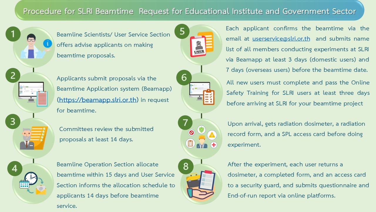
Beamline Submission Process
All procedures can be found in the following document. Click to download
Failure to submit the End of Experiment Feedback and/or the Experimental Report within the specified timeframe will be taken into consideration during the review of future beamtime proposals. Proposals may not be approved if the required reports from previous experiments have not been submitted.