Blog

  • Made in Thailand: A 7,000-Volt Power Supply Opening the Door to the Atomic-Scale Vacuum World

    Made in Thailand: A 7,000-Volt Power Supply Opening the Door to the Atomic-Scale Vacuum World

    Inside a synchrotron light source, tiny electrons cannot travel through ordinary atmospheric conditions. Instead, they must move within specially designed vacuum chambers that simulate conditions close to outer space. Ion pumps function like “mechanical lungs,” continuously removing air molecules from the vacuum chamber to maintain the ultra-clean environment required for electron circulation.

    To operate these pumps, a high-voltage power supply for ion pumps (Ion Pump Power Supply) is essential.

    Typically, the high-voltage power supplies used to control these pumps—operating at +7,000 V and −7,000 V—must be imported from abroad at extremely high cost. Recognizing this challenge, Thai engineers began developing a “Made in Thailand” high-voltage power supply capable of meeting international standards.

    The newly developed system offers several advanced features:

    • Performance comparable to internationally imported equipment
    • Touchscreen control interface with real-time status monitoring
    • Ethernet connectivity for remote operation and monitoring
    • Built-in interlock safety system that automatically cuts off power in case of abnormal conditions
    • Successfully tested in synchrotron accelerator systems including the Linear Beam Transport (LBT), Booster Synchrotron, and Storage Ring
    • Capable of supporting ultra-high vacuum (UHV) levels below 5 × 10⁻¹⁰ Torr

    Through the dedication and innovation of engineers at the Synchrotron Light Research Institute, this development represents an important milestone for Thailand’s vacuum technology sector.

    The innovation contributes to several long-term benefits:

    • Reducing reliance on imported technology – eliminating long procurement times and high costs
    • Supporting the development of advanced scientific instruments – including particle accelerators and advanced materials research laboratories, while paving the way for Thailand’s future 3 GeV synchrotron light source
    • Strengthening economic competitiveness – enabling technology transfer to Thai industries and creating potential for future export-oriented manufacturing

    With extensive experience in advanced vacuum technologies, the institute also provides expertise in designing, constructing, and maintaining integrated vacuum systems for organizations interested in adopting such technologies.

    Fun Fact:

    The 7,000-volt power used in this system is about 30 times higher than household electricity, yet it is carefully engineered to remain safe and stable while maintaining vacuum conditions as rarefied as outer space.

    This innovation is not merely a power supply—it is a key that unlocks the potential of Thai science on the global stage.

    —————————————————–

    Article by:

    Mr. Supan Boonsuya, Accelerator Maintenance and Operation Officer (Electrical Engineer)
  • “Miraculous Copper in Vacuum Chambers: An Innovation for Reducing Electron Beam Disturbances in Synchrotron Light Sources”

    “Miraculous Copper in Vacuum Chambers: An Innovation for Reducing Electron Beam Disturbances in Synchrotron Light Sources”

    A synchrotron light source—often described as a “giant microscope” used to study matter at the atomic scale—is facing a new technical challenge. High-energy electrons traveling through vacuum chambers at speeds close to that of light generate electromagnetic disturbances, which can affect the stability of the electron beam.

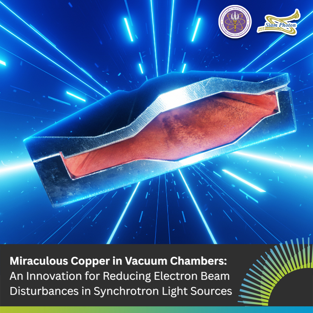
    To address this issue, researchers have developed a solution by coating the inner surface of the vacuum chamber with copper through an electroplating process. The copper layer must be extremely thin and uniformly deposited to enhance electrical conductivity and reduce energy losses.

    Why Copper Coating?

    Vacuum chambers are traditionally made from stainless steel. Although stainless steel is mechanically strong, it has relatively poor electrical conductivity. As high-energy electrons pass through the chamber, electromagnetic effects related to surface impedance can disturb the circulating electron beam. By coating the inner surface of the chamber with copper—an excellent electrical conductor—researchers effectively create a smooth, highly conductive pathway that allows electrons to travel more efficiently, minimizing interference along their path.

    Challenges in Electroplating Inside Vacuum Chambers

    Electroplating copper inside vacuum chambers requires precise engineering and strict control:

    • The copper layer must be thin yet highly uniform, so that it does not obstruct the electron beam path.
    • The surface must be extremely smooth to prevent unwanted electromagnetic reflections.
    • The coating must withstand high vacuum conditions and elevated temperatures during operation.

    Broader Impact of the Innovation

    • Reducing reliance on imported technologies
    • Strengthening the capabilities of Thai researchers and engineers in advanced vacuum component manufacturing
    • Supporting future development of particle accelerators and other scientific instruments

    Looking Ahead

    This development marks an important step toward high-performance vacuum chambers designed and manufactured in Thailand. By lowering costs and strengthening domestic expertise, it paves the way for further advancements in high-level scientific research.

    Fun Fact: 

    The copper coating inside the vacuum chamber is much thinner than human hair, yet it plays a role as crucial as a circulatory system in a synchrotron light source.

    ———————————————————

    Article by:

    Acting Sub Lt. Sarawut Chitthaisong, Accelerator Maintenance and Operation Officer

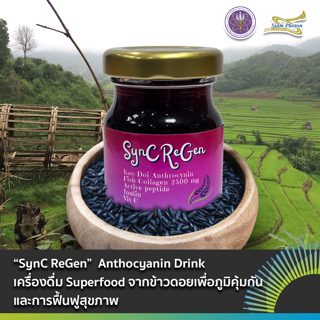
  • “SynC ReGen” Anthocyanin Drink Superfood Drink from Upland Rice for Immunity and Rehabilitation

    “SynC ReGen” Anthocyanin Drink Superfood Drink from Upland Rice for Immunity and Rehabilitation

    Nowadays, people around the world are increasingly paying attention to their health—especially following the COVID-19 pandemic—health food products have experienced rapid growth in popularity. The global health food market reached a value of USD 712.25 billion in 2024 and is projected to expand to USD 1,258.5 billion by 2030, with an average annual growth rate (CAGR) of approximately 10% during 2024–2030. Among the trending product categories is superfood, which refers to foods rich in concentrated nutrients such as vitamins, minerals, dietary fiber, antioxidants, and phytonutrients. These nutrients help strengthen the immune system, reduce the risk of chronic diseases, and support the body’s recovery and overall well-being.

    Highland Rice: A Unique Ingredient from Thailand’s Ethnic Communities

    Rice 3

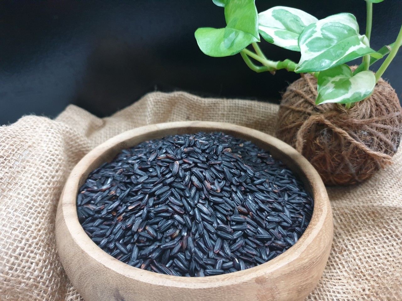
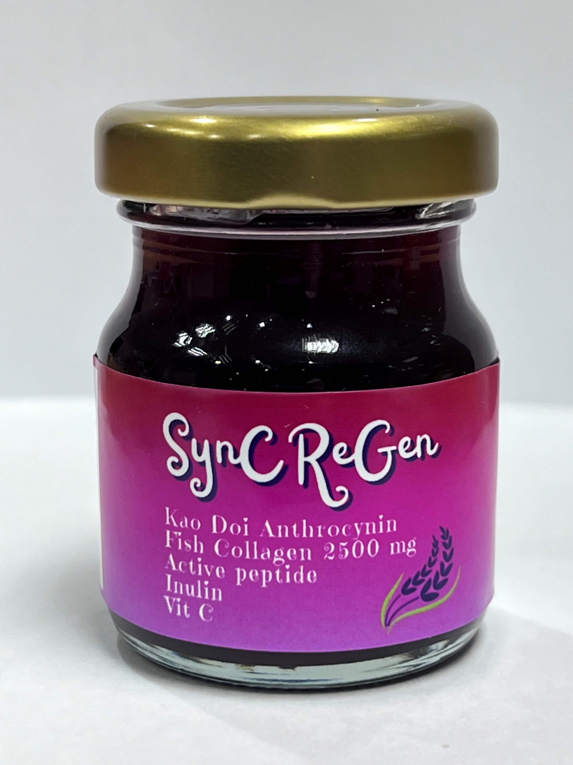
    Synchrotron Light Research Institute (SLRI) has developed a superfood beverage product derived from “Doi rice”, specifically the Jakunane variety, selected by the Highland Research and Development Institute (Public Organization). This indigenous rice variety is cultivated by ethnic communities living in the highland regions of northern Thailand. The rice grains have a deep purple color and are rich in minerals and phytochemicals, particularly anthocyanins and flavonoids, which possess strong antioxidant properties and contribute to immune system enhancement.

    In addition to highland rice extract, the beverage contains red dragon fruit extract, which is rich in Betalains, known for their antioxidant activity and high dietary fiber content. The product also includes inulin, a prebiotic that serves as food for beneficial gut bacteria and helps maintain a balanced intestinal microbiome. Furthermore, collagen derived from deep-sea fish supports cellular regeneration, while short-chain peptides and vitamin C are added to stimulate immune responses and promote cellular recovery.

    messageImage 1759995769470
    messageImage 1759996623287
    Picture1

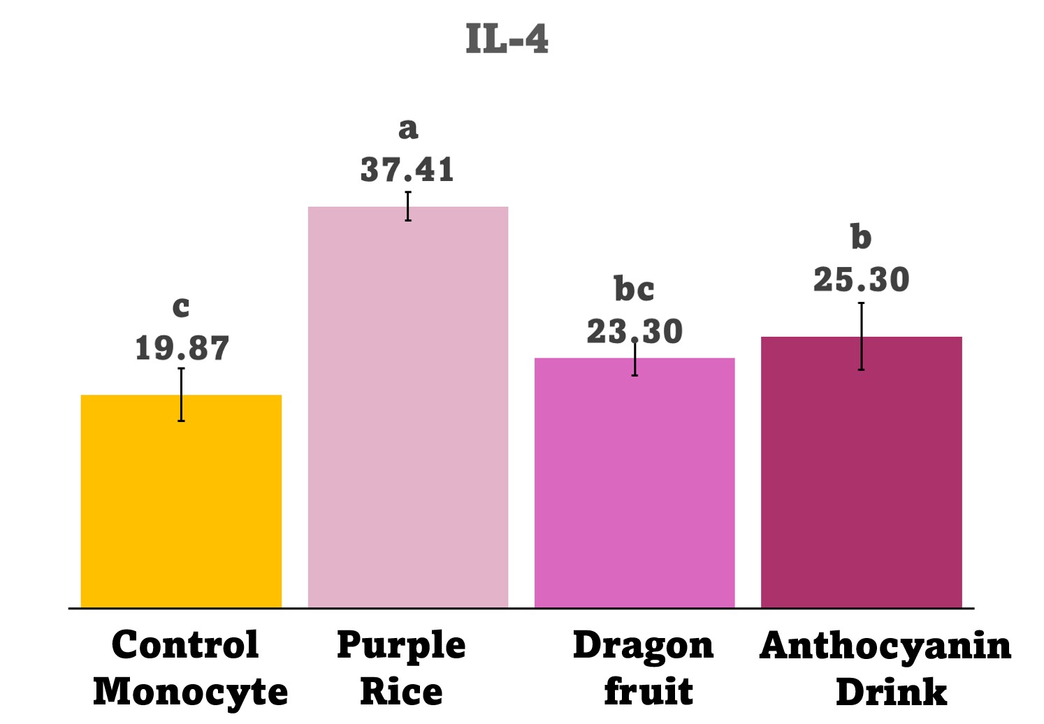
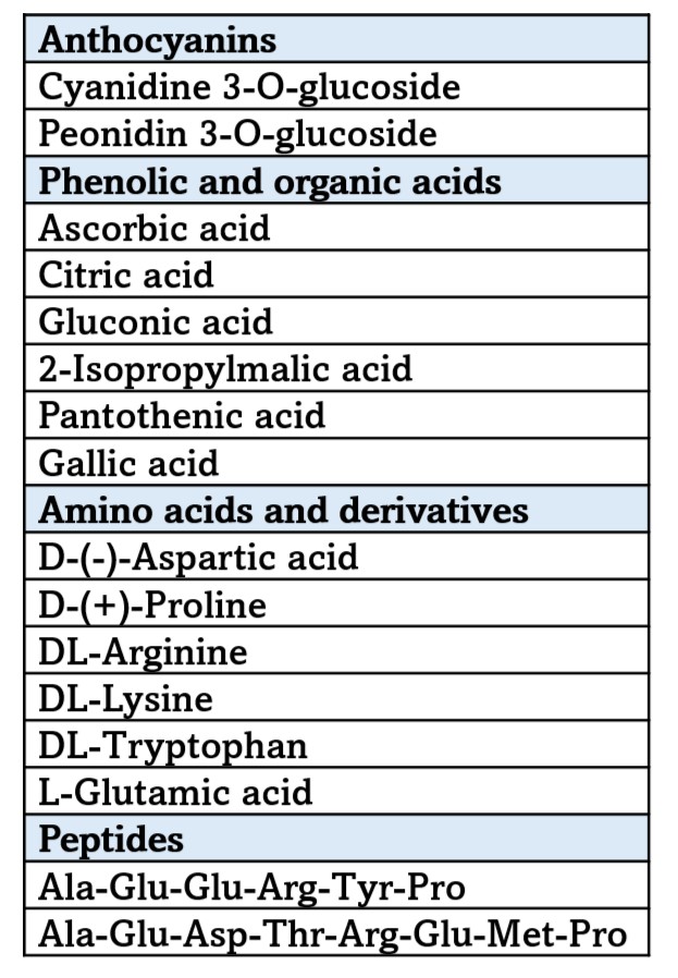
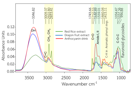
    Analytical studies conducted using advanced instruments at SLRI—including FTIR, LC-HRMS, XRF, and Tiny TIM—confirmed the presence and stability of key bioactive compounds in the product. The results demonstrated high antioxidant activity and immune-stimulating potential, while collagen absorption efficiency was found to exceed 88%. In addition, the product has passed safety and quality tests, with a shelf life of up to 90 days when stored at 4 °C.

    The beverage product “SynC ReGen” is not only a health drink but also an innovative superfood beverage that adds value to Thailand’s natural ingredients. Developed through scientific research and product innovation, it elevates local agricultural resources to an international standard while meeting the needs of consumers of all ages—from health-conscious individuals and working professionals to patients in recovery who seek comprehensive health support.

    00

    —————————————————

    Article by Dr. Waraporn Tanthanuch (Beamline Scientist, SLRI)

    Notice:

    Research Team

    Synchrotron Light Research Institute (Public Organization)

    Dr.Waraporn Tanthanuch, Dr.Sukanya Tastub, Dr.Buabarn Kuaprasert, Dr.Somchai Tancharakorn, Dr.Bussayarat Maikhunthod

    Highland Research and Development Institute (Public Organization)

    Dr.Janjira Rungcharoen

  • In Vivo Studying of P, S, K and Ca Distribution in Fungi Injured Soybean Leaves by µXRF

    In Vivo Studying of P, S, K and Ca Distribution in Fungi Injured Soybean Leaves by µXRF

    Micro X-ray fluorescence (µXRF, micro XRF) or XRF imaging is a powerful, non-destructive multielemental technique for the in vivo identification of plant tissues. It offers details of elemental distribution, contamination and nutrient absorption in plant tissues (Mongkhonsin et al., 2016; Rodrigues et al., 2018; Bonto et al., 2020; Nakhforoosh et al., 2024; Navarro et al., 2019; Vives et al., 2005).

    Micro XRF measurement does not require vacuum condition, thus being a practical tool for the direct analysis of fresh and in vivo plants, whereas laser-induced breakdown spectroscopy (LIBS) and laser ablation inductively coupled plasma mass spectrometry/optical emission spectroscopy (LA-ICP-MS/OES) are destructive techniques.

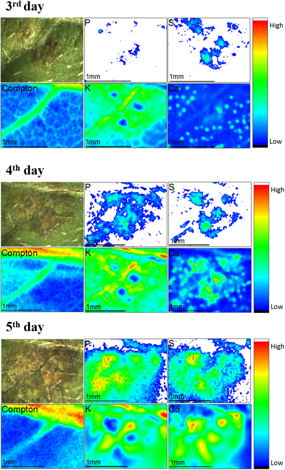
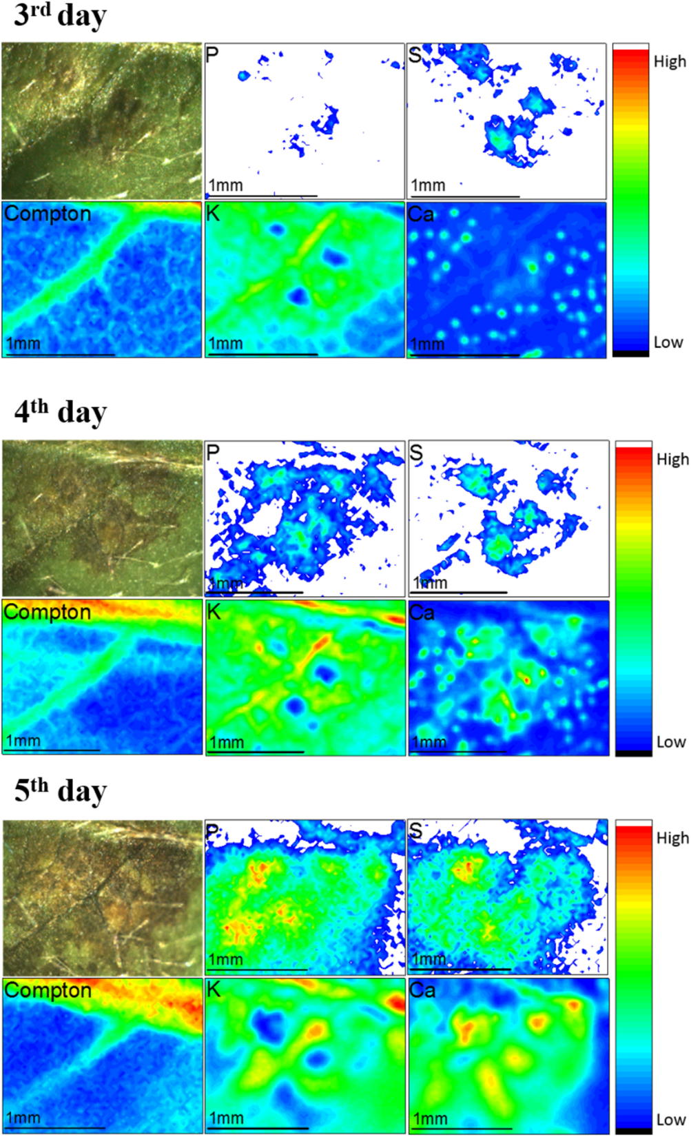
    Elemental distribution of soybean leaves infected by anthracnose disease is in vivo characterized by µXRF (Rodrigues et al., 2018). Micro XRF experiment provides elemental images of P, S, K and Ca in the infected soybean leaf from the 3rd to the 5th day after the fungi inoculation. Moreover, the presence of veins in the leaf was observed by the Compton scattering map.

    P, S, and Ca were accumulated in the injured region where the disease spreads. On the other hand, the micro XRF imaging resulted in lesser amount of K in the infected areas, thus corresponding to the darker areas.

    Micro XRF image of the soybean leaf infected by anthracnose disease from the 3rd, 4th and 5th day after fungus Colletotrichum truncatum inoculation is shown in Figure 1.

    In conclusion, the spread of diseases caused by fungus Colletotrichum truncatum significantly affected the elemental distribution in infected soybeans. High concentration of Ca in infected regions may relate to the influx of calcium ions to cells, thus being a trigger defense mechanism by the elicitors and plant proteins. Therefore, elemental accumulation may link with the self-defense mechanism of plants.

    Picture1 Plant Science

    Figure 1: Micro XRF elemental mapping of the soybean leaf infected by anthracnose disease from the 3rd, 4th and 5th day after fungus Colletotrichum truncatum inoculation

    Citation: Frontiers in Plant Science. 9, DOI: 10.3389/fpls.2018.01588.

    —————————————————

    Article by Chomphunuch Songsiriritthigul (Beamline Scientist, SLRI)

    References

    Bonto, A., Jearanaikoon, N., Sreenivasulu, N., Camacho, D. (2020). High uptake and inward diffusion of iron fortificant in ultrasonicated milled rice. LWT-Food Science and Technology. 128(5), 109459. DOI: 10.1016/j.lwt.2020.109459.

    Mongkhonsin, B., Nakbanpote, W., Hokura, A., Nuengchamnong, N., Maneechai, S. (2016). Phenolic compounds responding to zinc and/or cadmium treatments in Gynura pseudochina (L.) DC. extracts and biomass. Plant Physiology and Biochemistry. 10, 549-560. DOI: 10.1016/j.plaphy.2016.10.027.

    Nakhforoosh, A., Hallin, E., Karunakaran, C., Korbas, M., Stobbs, J., Kochian, L. (2024). Visualization and Quantitative Evaluation of Functional Structures of Soybean Root Nodules via Synchrotron X-ray Imaging. Plant Phenomics. 6, 0203. DOI: 10.34133/plantphenomics.0203.

    Navarro, H., Marcó, L. M., Araneda, A. A., & Bennun, L. (2019). Spatial distribution of Si in Pinus Insigne (Pinus radiata) Wood using micro XRF by Synchrotron Radiation. Journal of Wood Chemistry and Technology, 39(3), 187–197. DOI: 10.1080/02773813.2018.1562473.

    Rodrigues, E. S., Gomes, M. H. F., Duran, N. M., Cassanji, J. G. B., da Cruz, T. N. M., Sant’Anna Neto, A., Savassa, S. M., de Almeida, E., Carvalho, H. W. P. (2018). Laboratory Microprobe X-Ray Fluorescence in Plant Science: Emerging Applications and Case Studies. Frontiers in Plant Science. 9, DOI: 10.3389/fpls.2018.01588.

    Vives, A. E. S., Silva, R. M. C., Medeiros, J. G. da S., Tomazello Filho, M., Barroso, R. C., Zucchi, O. L. A. D., & Moreira, S. (2005). Accumulation of elements in annual tree rings measured by synchrotron x‐ray fluorescence analysis. X-Ray Spectrometry, 34(5), 411–416. DOI: 10.1002/xrs.844.

  • A Potential of Bioactive Compound as an Antitubercular Agent

    A Potential of Bioactive Compound as an Antitubercular Agent

    The disease tuberculosis (TB) which is caused by the bacterium Mycobacterium Tuberculosis, is an infectious disease listed among the top 10 causes of fatality worldwide. It killed up to 1.4 million people in 2019 (WHO), thus making it as one of the top 10 causes of death.

    The enoyl-acyl carrier protein reductase (InhA) from M. Tuberculosis, is one of the key enzymes which is responsible for synthesis of essential mycolic acids in the mycobacterial cell wall. Therefore, InhA is the key target for anti-tubercular drug. Multi drug resistant TB remains a public health crisis, thereafter, the studies of potent and novel inhibitors are of great interest.

    Therefore, Assoc. Prof. Dr. Pornpan Pungpo (Department of Chemistry, Faculty of Science, Ubon Ratchathani University) has been interested in the development of the potent and novel inhibitors.

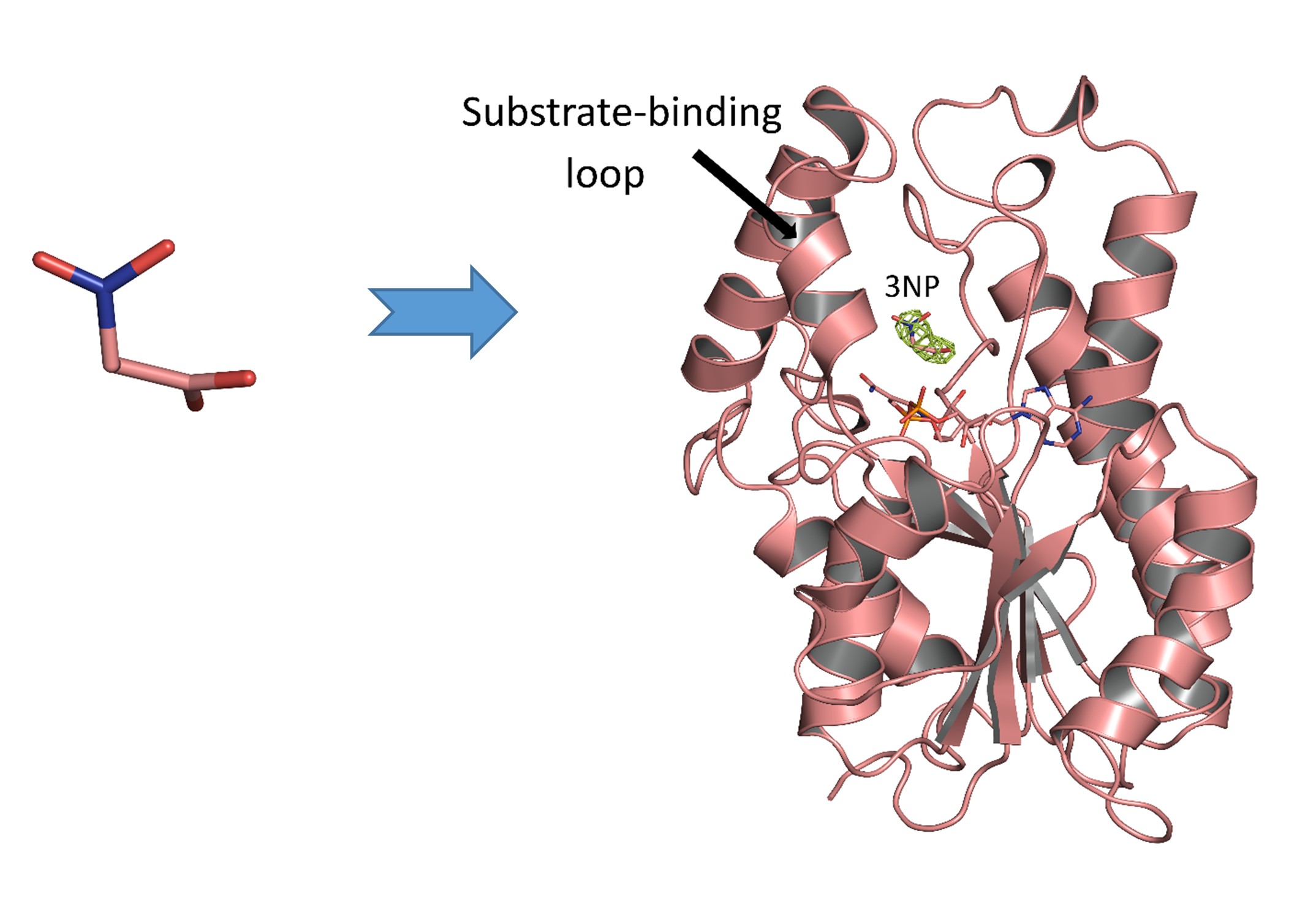
    3NP (Figure 1A) has been extracted from endophytic fungi, Phomopsis sp. usia5 from plant hosts located in Thailand. It is a bioactive compound and proposed as an antitubercular agent. It is a promising antimycobacterial agent, thus exhibiting minimum inhibitory concentration (MIC) values of 50.0 µg/mL and 12.5 µg/mL against M. tuberculosis H37Ra and M. tuberculosis H37Rv, respectively. 3NP showed half maximal inhibitory concentration (IC50) values for InhA inhibition of 71.2 μM, compared to a value for InhA inhibitor triclosan of 5.32 μM.

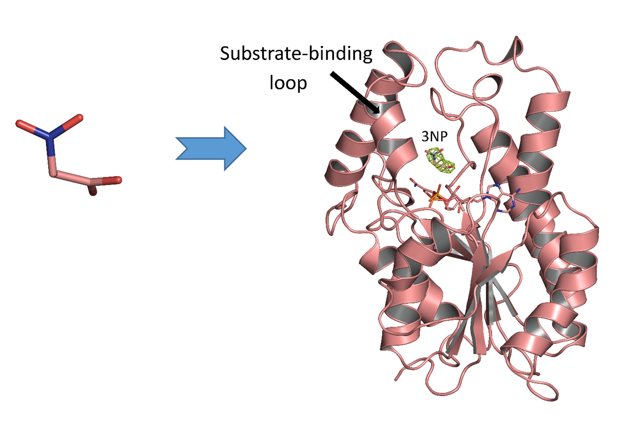
    Macromolecular Crystallography (MX) has been carried out for studying the three dimensional structure of holo structure of InhA from M. Tuberculosis (Figure 1B). Dr. Chomphunuch Songsiriritthigul (Synchrotron Light Research Institute, SLRI) performed crystal structure analysis under the supervision of Prof. Dr. James Spencer (School of Cellular and Molecular Medicine, University of Bristol, United Kingdom).

                                                                           (A)                                                              (B)

    Picture 3NP

    Figure 1 (A) Chemical structure of  3-nitropropanoic acid (3NP), a bioactive compound. (B) The overall structure of InhA-NAD+-3NP complex (PDB entry 7E48).

    Citation:  PROTEINS: Structure, Function, and Bioinformatics. 2022; 90(3): 898-904. DOI: 10.1002/prot.26268.

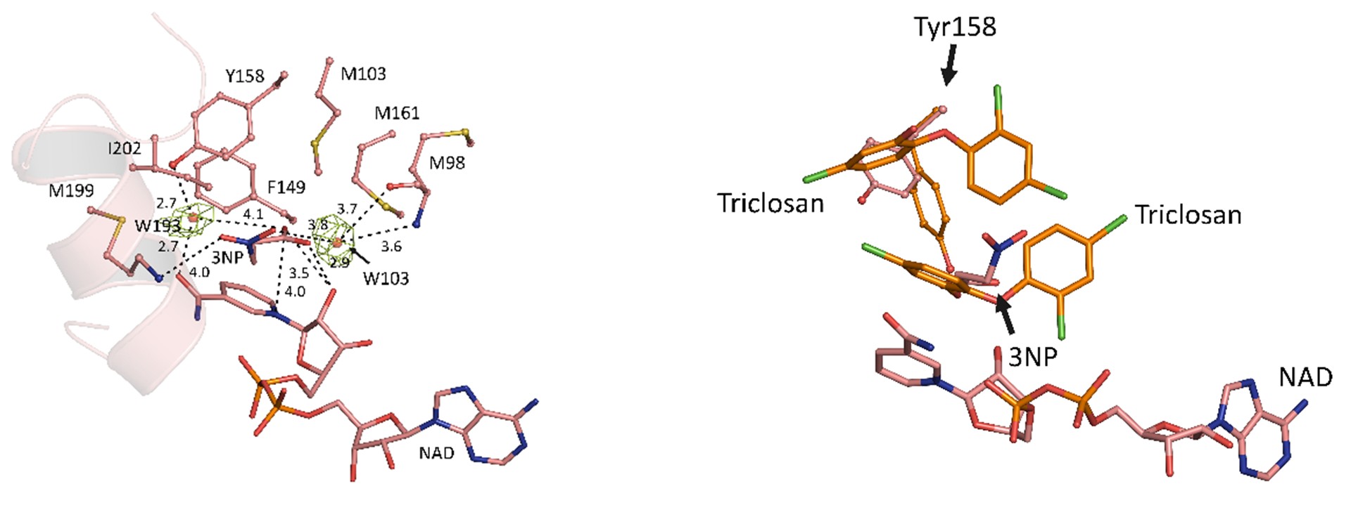
    Crystal structure of the complex contains the InhA substrate-binding loop in an ordered, open conformation with Tyr158, a catalytically important residue whose orientation defines different InhA substrate/inhibitor complexes, in the “out” position (Figure 2A). 3NP occupies a hydrophobic binding site adjacent to the NAD+ cofactor and close to that utilized by the diphenyl ether triclosan (Figure 2B), but binds predominantly via electrostatic and water-mediated hydrogen-bonding interaction with the protein backbone and NAD+ cofactor. 

                                                                           (A)                                                                  (B)

    Picture2 3NP

    Figure 2 (A) The structural components of InhA-NAD+-3NP complex (PDB entry 7E48, salmon). Water-mediated hydrogen bonds which stabilized both 3NP and NAD+ are shown. (B) The superposed structures of InhA-NAD+-3NP (PDB entry 7E48, salmon) and InhA-NAD+-triclosan (PDB entry 1P45, orange).

    Citation:  PROTEINS: Structure, Function, and Bioinformatics. 2022; 90(3): 898-904. DOI: 10.1002/prot.26268.

    Therefore, 3NP retains value as a starting point as a pharmacophore for potential antituberculosis drugs owing to its ready availability from natural and commercial sources, the potential for developing derivatives that may reduce toxicity and improve InhA inhibitory activity.

    The lack of involvement of specific amino acid side chains in 3NP binding suggests that compounds exploiting these interactions might show reduced susceptibility to emergence of resistance by mutation of the InhA active site. These data indicate that further exploration and compound optimization, for example derivatization of 3NP, may be warranted to exploit these findings in potential antituberculosis agents. 

    ———————————————————————————————-

    Written by Chomphunuch Songsiriritthigul (Beamline Scientist, SLRI)

    References

    1. Chomcheon, P, Wiyakrutta, S, Sriubolmas, N, Ngamrojanavanich, N, Isarangkul, D, Kittakoop, P. 3-Nitropropionic Acid (3-NPA), a Potent Antimycobacterial Agent from Endophytic Fungi: Is 3-NPA in Some Plants Produced by Endophytes?. J Nat Prod. 2005; 68: 1103-1105.
    2. Ganihigama, DU, Sureram, S, Sangher, S, Hongmanee, P, Aree, T, Mahidol, C, Ruchirawat, S, Kittakoop, P. Antimycobacterial activity of natural products and synthetic agents: Pyrrolodiquinolines and vermelhotin as anti-tubercular leads against clinical multidrug resistant isolates of Mycobacterium tuberculosis. Eur J Med Chem. 2015; 89: 1-12.
    3. Kamsri, P, Hanwarinroj, C, Naruedon, P, et al. Discovery of New and Potent InhA Inhibitors as Antituberculosis Agents: Structure-Based Virtual Screening Validated by Biological Assays and X‑ray Crystallography. J Chem Inf Model. 2019; 60: 226-234.
    4. Songsiriritthigul, C., Hanwarinroj, C., Pakamwong, B., Srimanote, P., Suttipanta, N., Sureram, S., Suttisintong, K., Kamsri, P., Punkvang, A., Spencer, J., Kittakoop, P., Pungpo, P. Inhibition of Mycobacterium tuberculosis InhA by 3-nitropropanoic acid. Proteins: Struct Funct Bioinf. 2022; 90(3): 898-904. DOI: 10.1002/prot.26268.
    5. World Health Organization. https://www.who.int/news-room/fact-sheets/detail/tuberculosis. Accessed: February 2021.
  • Synchrotron X-ray Fluorescence Analysis of Functional Polysaccharides Derived from Spirulina

    Synchrotron X-ray Fluorescence Analysis of Functional Polysaccharides Derived from Spirulina

    Spirulina, a type of microalga, is well-known for its high nutritional content and various bioactive compounds. In particular, polysaccharide extracts from Spirulina have garnered interest due to their biological properties and numerous health benefits.

    Dr. Wanida Pan-utai from the Department of Applied Microbiology at the Institute of Food Research and Product Development, Kasetsart University, and her research team have optimized a protocol for extracting crude polysaccharides using water extraction. They have also characterized the biological properties of these polysaccharides.

    Polysaccharide extracts from oven-dried biomass of Spirulina exhibited promising antioxidant properties, as determined through three different assays: the DPPH Assay, ABTS Assay, and FRAP Assay. Furthermore, the extract demonstrated a greater suppression of alpha-glucosidase activity compared to the original oven-dried biomass, making it a potential option for controlling blood glucose levels and a safer alternative with reduced chemical use.

    The growth of probiotics from the polysaccharide powder was found to be higher than that from inulin, a commercial prebiotic known for stimulating the growth of beneficial bacteria.

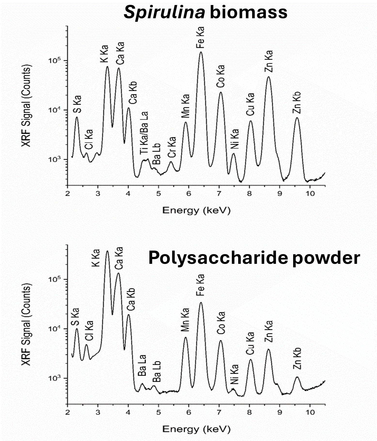
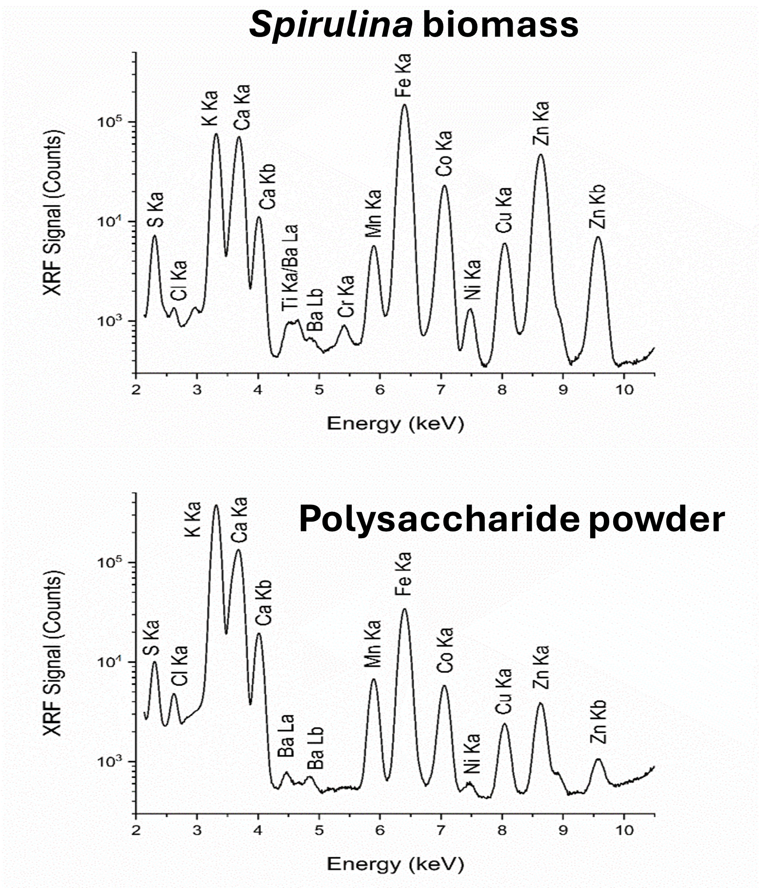
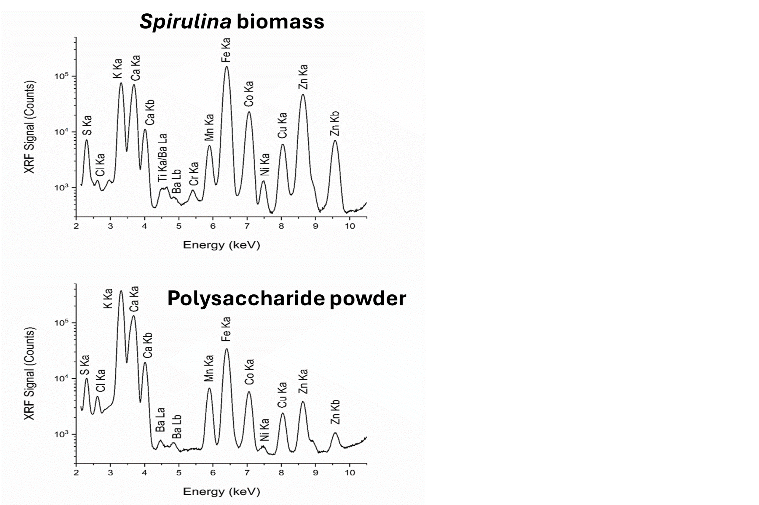
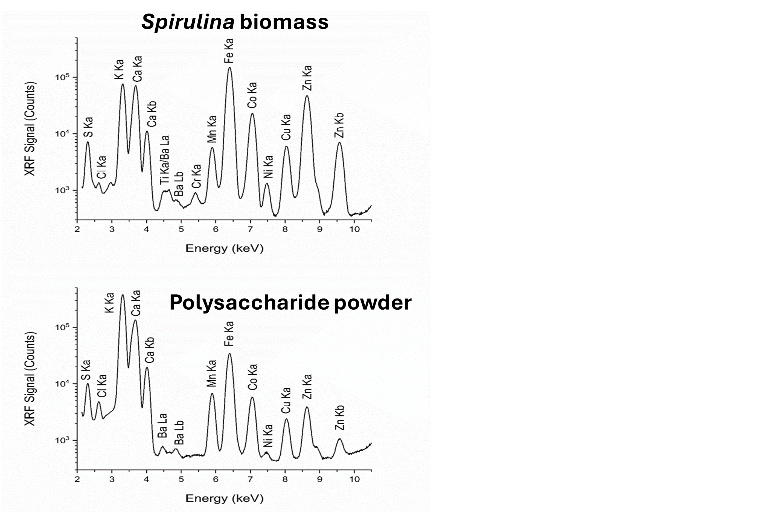
    The elemental compositions of the extract were analyzed using the synchrotron X-ray fluorescence (XRF) technique at Beamline 7.2W of the Synchrotron Light Research Institute (SLRI) in Thailand. Figure 1 presents the X-ray fluorescence spectrum of both the Spirulina biomass and the polysaccharide powder derived from it.

    Figure 1

    Figure 1 Synchrotron X-ray fluorescence spectra of Spirulina biomass and polysaccharide powder derived from Spirulina

    Citation: Life. 15, 1213. DOI: 10.3390/life15081213.

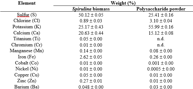
    Table 1 Weight percentages from X-ray fluorescence (XRF) analysis.

    Screenshot 2025 09 30 083438

    Values are presented as mean ± standard deviation (SD), where “n.d.” denotes not detected.

    Citation: Life. 15, 1213. DOI: 10.3390/life15081213.

    The X-ray fluorescence (XRF) technique was used to simultaneously detect various elements in the samples, reporting their weight percentages. In the polysaccharide powder, the predominant inorganic mineral nutrients identified were sulfur (S), chlorine (Cl), potassium (K), and calcium (Ca), while iron (Fe) was found to be a minor element. Additionally, trace elements such as manganese (Mn), cobalt (Co), nickel (Ni), copper (Cu), zinc (Zn), and barium (Ba) were detected in the extractant.

    This highlights the significant advantage of utilizing beamline capabilities to identify inorganic mineral nutrients, even when they are present in trace amounts. Furthermore, employing the synchrotron XRF technique in this research enables the simultaneous detection of a wide range of trace elements without the need for any chemical pretreatment, thereby reflecting their nutritive properties.

    ————————————–

    Article by Chomphunuch Songsiriritthigul (Beamline Scientist, SLRI)

    References

    Anwer, S. S. (2023). Direct analysis of algae for determination of major and minor elements using energy dispersive x-ray fluorescence (EDXRF). Kuwait Journal of Science, 50(4): 697–702. DOI: 10.1016/j.kjs.2023.04.012.

    Pan-utai, W., Phomkaivon, N., Settachaimonkon, S., Pongponpai, P., Songsiriritthigul, C. (2025). Extraction, Characterization, Biological Properties, and X-ray Fluorescence Analysis of Functional Polysaccharides Derived from Limnospira platensis. Life. 15, 1213. DOI: 10.3390/life15081213.

  • Unveiling Ruby Origins with Synchrotron XANES

    Unveiling Ruby Origins with Synchrotron XANES

    The color of rubies, which are scientifically classified as corundum (Al₂O₃), is mainly determined by the presence and oxidation states of trace metals such as chromium, iron, and titanium. Understanding these impurities is essential for gemologists, as it enables them to assess the aesthetic value of gemstones and identify their geographic origins (Bootchanont et al., 2022, Bootkul et al., 2025, Intarasiri et al., 2016).

    A powerful technique for examining the oxidation states of trace elements is X-ray Absorption Near Edge Structure (XANES) spectroscopy. Recently, Dr. Saweat Intarasiri and his team at the Multidisciplinary Research Institute at Chiang Mai University have utilized this method to study rubies (Bootkul et al., 2025). Their research combines Laser Ablation Inductively Coupled Plasma Mass Spectrometry (LA-ICP-MS) with synchrotron-based XANES to provide a more comprehensive understanding of trace element composition and help identify their origins.

    Chromium (Cr): The Red Signature of Ruby

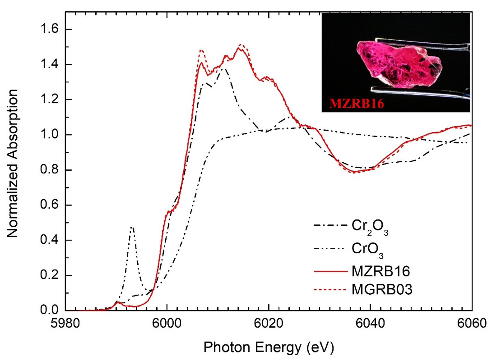
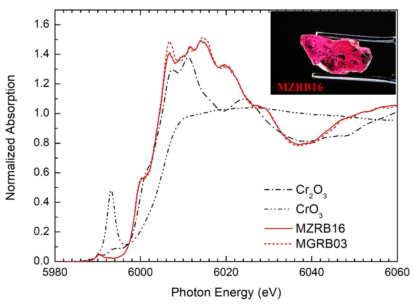
    Chromium ions are responsible for the characteristic red hues of rubies, which can range from pale pink to deep crimson depending on their concentration. In this study, we compared the Cr K-edge XANES spectra of reference standards—Cr₂O₃ for Cr³⁺ and CrO₃ for Cr⁶⁺—with natural ruby samples from Mozambique (MZRB16) and Madagascar (MGRB03). The measurements were conducted at Beamline 7.2W of the Synchrotron Light Research Institute (SLRI). The results confirmed that chromium in both ruby samples exists predominantly in the +3 oxidation state (Cr³⁺), which is a common characteristic of natural rubies worldwide (Bootkul et al., 2025).

    Figure 1 Cr K-edge XANES spectra showing reference standards (Cr₂O₃, CrO₃) and natural ruby samples (MZRB16 and MGRB03) collected at Beamline 7.2W, Synchrotron Light Research Institute (SLRI).

    Citation: Spectrochimica Acta Part B: Atomic Spectroscopy, Vol. 232. DOI: 10.1016/j.sab.2025.107271

    Titanium (Ti): Clues to Geological Origins

    Titanium plays a significant role in the chemistry of rubies. A marble-hosted ruby from Mong Hsu, Myanmar (MMRB04), which contains a relatively high concentration of titanium, was analyzed using X-ray Absorption Near Edge Structure (XANES) spectroscopy (Bootkul et al., 2025). The spectral features observed, including the pre-edge, shoulder, and main prominent peak, closely resembled those of rutile (TiO₂). This similarity suggests that the titanium in the ruby is in a comparable chemical state. These findings could indicate unique geological conditions at the origin of the ruby.

    By combining synchrotron XANES with LA-ICP-MS, researchers can identify the oxidation states and distribution of trace metals such as Cr, Fe, and Ti. This approach improves our ability to accurately determine the provenance of rubies from Myanmar, Madagascar, and Mozambique (Bootkul et al., 2025).

    This work highlights how cutting-edge synchrotron techniques can support not only scientific inquiry but also practical applications in gemology, mineral exploration, and the preservation of cultural heritage (Bootkul et al., 2025).

    ——————

    Article by Chomphunuch Songsiriritthigul (Beamline Scientist, SLRI)

    References

    Bootchanont A., Wattanawikkam C., Porjai P., Sailuam W., Busayaporn W., Saiyasombat C., Kidkhunthod P., Borsup J., Songsiriritthigul P., Jiamprasertboon A., Lertvanithphol T., Horprathum M., Pengsri P., Saisopa T. (2022). Characterization of structural orientation and optical properties of Al and Cr in rubies. Radiation Physics and Chemistry, Vol. 199. DOI: 10.1016/j.radphyschem.2022.110315.

    Bootkul, D., Intayot, S., Uthaichana, K., Wattanachai, P., Intarasiri, S., Songsiriritthigul, C., Songsiriritthigul, P. Hauzenberger, C. A. (2025). Reliability of LA-ICP-MS and Synchrotron XANES for provenance identification of rubies. Spectrochimica Acta Part B: Atomic Spectroscopy, Vol. 232. DOI: 10.1016/j.sab.2025.107271

    Intarasiri S., Bootkul D., Tippawan U., Songsiriritthigul P. (2016). Color improvement of rubies by ion beam technique. Surface and Coatings Technology, Vol. 306. pp. 205-210, DOI: 10.1016/j.surfcoat.2016.05.083.

  • Synchrotron X-ray footprinting for studying the structure and dynamics of biomolecules

    Synchrotron X-ray footprinting for studying the structure and dynamics of biomolecules

               Synchrotron X-ray footprinting (XF or XFP) provides a new method for studying macromolecular structures in the native solution state. XFP technique employs broadband hard X-ray beams to generate hydroxyl radicals in solution, thus subsequently covalently modifying amino acid side chains of protein structure and cleaving the phosphodiester backbone of nucleic acids.

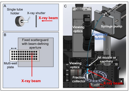
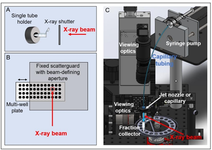
               High flux of synchrotron X-ray beams produce hydroxyl radicals for X-ray footprinting experiments to study structural information and conformational changes of proteins and nucleic acids. Up to now, XFP beamlines have been implemented to two national synchrotron facilities, the Advanced Light Source (ALS) and the National Synchrotron Light Source (NSLS) in USA. The XFP beamline of NSLS utilizes hard X-rays from a three-pole wiggler (3PW) source, thus delivering high flux at sample in the range of 1×10¹⁶ phs/s. Figure 1 illustrates the XFP end station, showing various sample delivering systems. X-ray shutter is employed for exposure of sample solution which is contained in the tube. The duration of X-ray exposure much depends on the velocity of the sample passing the X-ray beam in multi-well plate setup and jet nozzle, or capillary.

    nmzbfdhbzx
    Figure 1 Sample delivery system at the end station of XFP beamlines. (A) Single tube holder (B) Multi-well plate (C) Jet nozzle or capillary (Ralston & Joshua, 2022).

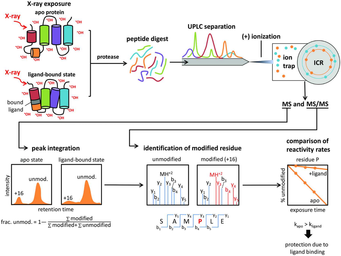
    A schematic diagram of protein X-ray footprinting measurements is shown in Figure 2. An X-ray beam exposes to apo and ligand-bound (holo) proteins, thus subsequently digesting with proteases and generating peptides, separating by Mass Spectrometry. The fragmentation pattern is unique to each peptide; therefore MS/MS spectra is employed to identify unmodified and modified peptides/residues within the sample. Peak identification of extracted ion chromatograms is for quantifying the fraction of the unmodified and modified peptides/residues within the sample and determining regions of the protein which are responsible for specific function.

    mdfgvbrye
    Figure 2. Schematic diagram of protein X-ray footprinting measurement. An X-ray beam exposed to apo and ligand-bound proteins. Digested peptides are separated by ultraperformance liquid chromatography technique (UPLC). Identification of the unmodified and modified peptides/residues is analyzed by electrospray ionization coupled with tandem mass spectrometry (ESI-MS/MS) (Asuru et al., 2019).

               Recently, XFP is an ideal complementary technique, thus comparing to low-to-medium resolution data from cryo-EM or solution scattering studies. Although crystallography and NMR provide high-resolution information. However, crystallization procedures and size limitations are much considered as bottlenecks. Static and time-resolved structural analysis of complex structures brings benefit of XFP technique, thus continuously developing to meet its highest performance.

               Re-written by Chomphunuch Songsiriritthigul (Beamline Scientist, SLRI)

    References

    1. Asuru, A., Farquhar, E.R., Sullivan, M., Abel, D., Toomey, J., Chance, M.R. & Bohon, J. (2019). The XFP (17-BM) beamline for X-ray footprinting at NSLS-II. Journal of Synchrotron Radiation. 26, 1388-1399. DOI: 10.1107/S1600577519003576.
    2. Chance, M.R., Farquhar, E.R., Yang, S., Lodowski, D.T. & Kiselar, J. (2020). Protein Footprinting: Auxiliary Engine to Power the Structural Biology Revolution. Journal of Molecular Biology. 432, 2973-2984. DOI: 10.1016/j.jmb.2020.02.011.
    3. Gupta, S. (2019). Using X-ray Footprinting and Mass Spectrometry to Study the Structure and Function of Membrane Proteins. Protein & Peptide Letters. 26(1):44-54. DOI: 10.2174/0929866526666181128142401.
    4. Morton, S.A., Gupta, S., Petzold, C.J. & Ralston, C.Y. (2019). Recent Advances in X-Ray Hydroxyl Radical Footprinting at the Advanced Light Source Synchrotron. Protein Peptide Letters. 26(1), 70-75. DOI: 10.2174/0929866526666181128125725.
    5. Ralston, C.Y. & Joshua S.S. (2022). Structural Investigation of Therapeutic Antibodies Using Hydroxyl Radical Protein Footprinting Methods. Antibodies11. 71. DOI: 10.3390/antib11040071.
  • Herbicides biodegradation by aldo-keto reductase enzyme

    Herbicides biodegradation by aldo-keto reductase enzyme

       Glyphosate, an organophosphorus compound, is the most extensively employed herbicide in agriculture worldwide including in Thailand, due to its efficient weed elimination. The widespread of glyphosate in water and soil is caused by inappropriate application practices and over-spraying herbicide, thus affecting the potential risks to human health. Moreover, the detection of glyphosate residues in human urine samples results in an increased exposure to glyphosate in humans. Currently, the selective analysis of glyphosate in environmental samples provides an early warning in incidents of contamination, thus being important for public health and food security.

               This research work investigated the capability of the aldo-keto reductases (AKRs) in glyphosate degradation, thus improving glyphosate potency in weed management. AKR Superfamily comprises enzymes that have broad substrate specificity and play specific roles in reactive aldehyde and ketone detoxification. In this study, AKRs, designated as AKR4C16 and AKR4C17, were found to contribute to glyphosate resistance. 

               The genes (EcAKR4–1 and EcAKR4–2) designated as AKR4C16 and AKR4C17, respectively in an AKR Superfamily demonstrated glyphosate degradation, thus suggesting the potential role in glyphosate detoxification. The control rice plants were killed by applied glyphosate at rates of 540 g ha-1 or higher as shown in Figure 1. However, the EcAKR4-1-overexpressing rice died at 2,160 g glyphosate ha-1 but it still survived at 540 and 1,080 glyphosate g ha-1 (Figure 1).

                Growth response to glyphosate was compared between the GFP (control) and EcAKR4-1 gene (Figure 1), therefore AKR4C16 confers glyphosate resistance in rice. In conclusion, the enzyme activity for glyphosate degradation was significantly higher in the transgenic lines expressing AKR4C16 compared to the wild type.

    Picture1

    Figure 1 The GFP (control) and transgenic rice seedlings after three weeks of glyphosate treatment. 

                  Crystal structures of AKR4C16 and AKR4C17 have been studied by the Macromolecular Crystallography technique. Refined structures of AKR4C16 and AKR4C17 adopt the same overall fold of a triosephosphate isomerase (TIM) motif. Their cofactor- and putative substrate-binding pockets are primarily identical. Therefore, AKR4C16 and AKR4C17 might exploit the same enzymatic mechanism to catalytically degrade glyphosate.

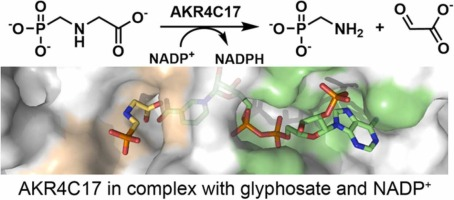
                  The catalytic process of glyphosate biodegradation is proposedly illustrated in Figure 2. The ternary complex structure reveals that both its cofactor and glyphosate are located at the long tunnel of the AKR4C17 structure as shown in Figure 2. The binding regions of NADP+ and glyphosate are displayed in green and yellow on the protein surface of AKR4C17, respectively.

    sdadse11

    Figure 2 The putative catalytic process and the cofactor and substrate-binding site of AKR4C17.

                Thereafter, the F291D mutant obtained from structure-based rational design enhances a 70% increase in enzyme activity for glyphosate degradation. Proposedly, the mutant structure preserved the overall fold and cofactor-binding features of wild-type enzymes. More hydrophilic interactions may assist glyphosate biodegradation, thus altering the electrostatic surface of the putative substrate-binding site.

                The dimensional structure of AKR4C17, an aldo-keto reductase could investigate its role in the glyphosate detoxification process, thus increasing the degree of glyphosate tolerance. Therefore, information obtained including the function and molecular structure of AKR4C147 might be very important in developing glyphosate-tolerant plants in the future.

    Re-written by Chomphunuch Songsiriritthigul (Beamline Scientist, SLRI)

    References

    1. Li, H., Yang, Y., Hu, Y., Chen, C-C., Huang, J-W., Min, J., Dai, L., Guo, R-T. (2022). Structural analysis and engineering of aldo-keto reductase from glyphosate-resistant Echinochloa colona. Journal of Hazardous Materials. DOI: 10.1016/j.jhazmat.2022.129191.
    2. Butmee, P., Tumcharern, G., Songsiriritthigul, C., Durand, M., J., Thouand, G., Kerr, M., Kalcher, K. & Samphao, A. (2021). Enzymatic electrochemical biosensor for glyphosate detection based on acid phosphatase inhibition. Analytical and Bioanalytical Chemistry. DOI: 10.1007/s00216-021-03567-2.
    3. Songsiriritthigul, C., Narawongsanont, R., Tantitadapitak, C., Guan, H-H. & Chen, C-J. (2020). Structure-function study of AKR4C14, an aldo-keto reductase from Thai Jasmine rice (Oryza sativa L. ssp. Indica cv. KDML105. Acta Crystallographica D. D76, 472-483.
    4. Pan, L., Yu, Q., Han, H., Mao, L., Nyporko, A., Fan, L., Bai, L., Powles, S. (2019). Aldo-keto reductase metabolizes glyphosate and confers glyphosate resistance in Echinochloa colona. Plant Physiology. 181 (4), 1519–1534. DOI: 10.1104/pp.19.00979.