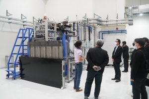
Welding high vacuum parts is knowledge developed by the Synchrotron Light Research Institute, as these welded samples are essential equipment for accelerators and beamlines. Therefore, the features and quality of the samples must be inspected to evaluate the performance of the vacuum materials produced.
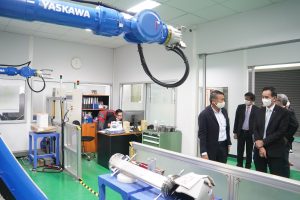
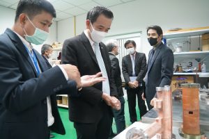
An example of welding vacuum materials at SLRI’s “Instrument Fabrication Section” which we designed and developed, is the welding of austenitic stainless steel AISI304 samples with oxygen-free copper (OFHC copper) using the Gas Tungsten Arc Welding process. This process produces sample materials with the ability to transfer heat and are strong, making them suitable for using in various devices in accelerators and beamline systems, such as photon beam masks and photon beam slits.
These vacuum materials are fabricated by welding together different metals. Scientists brought the samples to beamline 7.2W: Macromolecular Crystallography (MX) for feature and quality checks, using hard X-ray synchrotron light from the Macromolecular Crystallography beamline, obtained from a 6.5 Tesla superconducting wavelength shifter. The light is focused with an X-ray polycapillary half lens, resulting in a focal point of 42 microns, sufficient for distinguishing each area of the welded part.
Scientists employed micro-X-ray fluorescence spectroscopy (XRF) and micro-X-ray absorption spectroscopy (XAS) technique to study the welded AISI304 stainless steel and copper, as these techniques are suitable for examining the type of element and its composition in small areas on the nonhomogeneous sample.
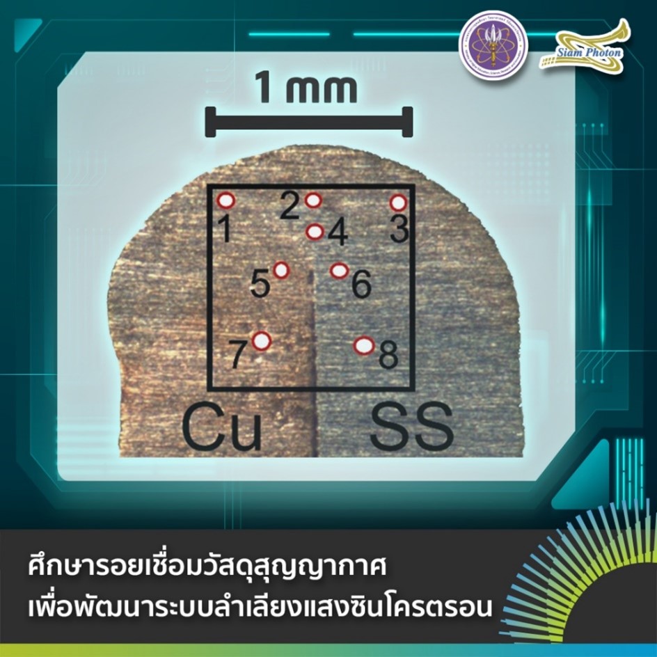
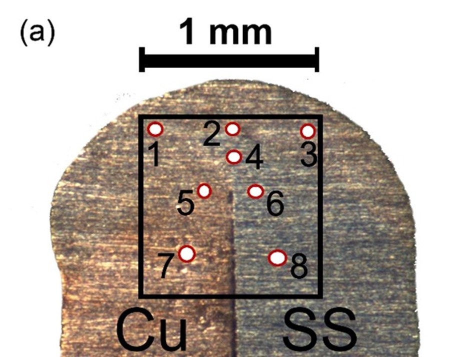
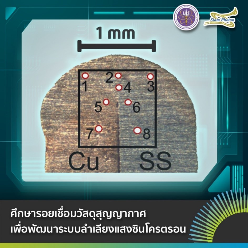
(Figure a) Sample welded between AISI304 stainless steel and copper with 8 analysis areas.
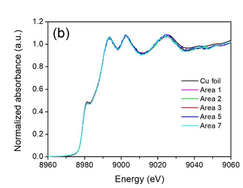
(Figure B) X-ray absorption spectra at the K-edge absorption region of various elements tested at Beamline 7.2, SLRI.
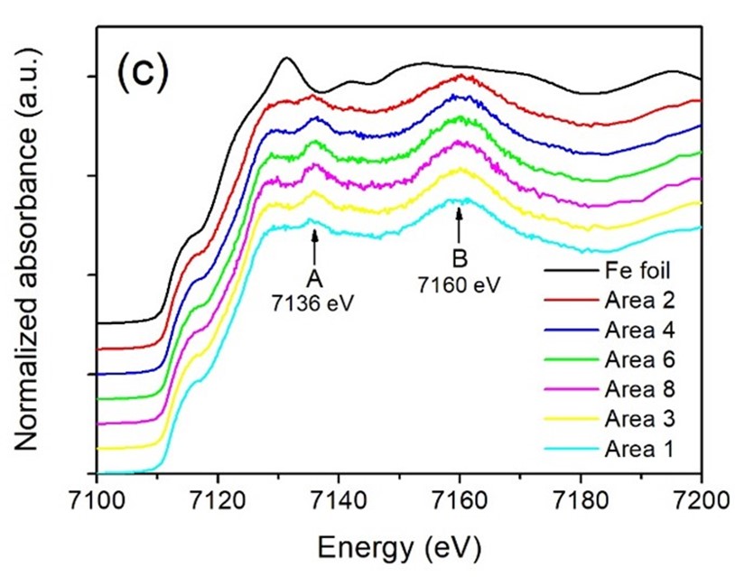
(Figure C) X-ray absorption spectra at the K-edge absorption region of various elements tested at Beamline 7.2, SLRI.
(Figure D) X-ray absorption spectra at the K-edge absorption region of various elements tested at Beamline 7.2, SLRI.
The examination of the sample from Figure A with synchrotron light at beamline 7.2W reveals the X-ray absorption spectrum at the K-edge absorption area of various elements, as depicted in Figures B, C, and D. The spectral characteristics of copper in various areas showed no difference (Figure B). However, the results exhibit a distinct pattern in the steel spectrum, as seen in Figure C, between the stainless-steel area (Area 8, SS side) and the welding joint area (Area 2), characterized by the presence of peaks A and B in the former.
The results indicate that Manganese, comprising 2 percent by weight, could replace iron atoms, exhibiting a structure resembling Face-Centered Cubic. This is evident in the stainless-steel area (Area 8, SS side) as shown in Figure D.
The sample examination is a result of continuous development and improvement of the beamline system, aimed at utilizing synchrotron light cost-effectively and efficiently. The high intensity X-ray focused to a small scale at the micrometer level is useful in distinguishing various features within small areas of a sample. Moreover, it is highly probable to be applied as a tool for analyzing more diverse types of samples in the future.
“Jaeng” is a plant that is spread around dry evergreen forests, mixed deciduous forests, dry dipterocarp forests, and limestone hills. The name in scientific term is Maerua siamensis (Kurz) Pax. The reason why it has “siam” in the name is to indicate that Thailand had discovered only one kind of Jaeng. Later, it was discovered by a team of researchers from Plant Genetic Conservation Project Under the Royal Initiative of Her Royal Highness Princess Maha Chakri Sirindhorn (RSPG), Suranaree University of Technology (SUT) in collaboration with the Nakhon Ratchasima Agricultural Research and Development Center, Nakhon Ratchasima Rajabhat University that Thailand also has another plant that has the same botanical characteristics as “Jaeng Siam”. We have discovered a new species of the world that is distributed only in the area of Sikhio District and Dan Khun Thot District, Nakhon Ratchasima Province.
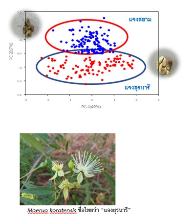
The new species of Jaeng has leaf morphology and appearance different from Jaeng Siam, but they have similar inflorescences and florets. This is an important morphological characteristic which indicates that they are plants of the same genus. Dr. Pratchaya Srisanga, a botanist from the Botanical Garden Organization and Assoc. Prof. Dr. Santi Watthana, botanist from SUT worked together to check the botanical name. They discovered the new species of the world in 2021 and named it in a botanical term as Maerea koratensis Srisanga & Watthana, and it is named in Thai as “Jaeng Suranaree” to remember Thao Suranaree and in honor of SUT. For the characteristics, Jaeng Suranaree is a semi-climbing shrub. It grows slowly in dry mixed forests. And is considered a local wild plant that is at risk of extinction.
Nevertheless, scientists have proven the uniqueness of the Jaeng Suranaree seed by using infrared radiation from synchrotron light, analyzed with the SR IR microspectroscopy technique by comparing the Jaeng Suranaree seed to the Jaeng Siam seed. The samples of Jaeng Suranaree and Jaeng Siam were collected from Lat Buakhao Subdistrict, Sikhio District, Nakhon Ratchasima Province. The result shows that the Jaeng Suranaree seed has a higher amount of fat than Jaeng Siam. However, Jaeng Siam seed has higher protein and carbohydrates than Jaeng Suranaree.
This basic information is the new knowledge that will help people to understand the basic information of Jaeng Suranaree. This will lead to in-depth studies, such as studies in other parts of Jaeng Suranaree including the study of various important active ingredients, such as determining the amount of total phenolic compounds and flavonoid compounds that are antioxidants or finding biologically active substances to develop into pharmaceuticals or cosmeceuticals, etc. It will further upgrade and develop Thai medicinal plants to have high added value.
Article by:
Assoc. Prof. Dr. Amornrat Molee, Director of the Plant Genetic Conservation Project Under the Royal Initiative of Her Royal Highness Princess Maha Chakri Sirindhorn, Suranaree University of Technology
Dr. Kanjana Thumanu, Beamline Scientist, Synchrotron Light Research Institute (Public Organization)
Complier: Corporate Communications Section, SLRI
Translation: International Relations Officer, User Service Section, SLRI
Currently, the market value of coffee around the world still tends to increase every year Even though Thailand can grow and produce both Arabica and Robusta coffee beans, however, the current amount of coffee production is still unable to compete with leading coffee exporting countries in the world market such as Brazil, Ethiopia, and Vietnam due to the limitation in planting areas in the country.
Therefore, an important factor in promoting the Thai coffee industry is to improve the quality of Thai coffee by creating the identity of Thai coffee, such as promoting the production of special coffee (Specialty coffee), which is high-quality coffee. It smells and tastes good. And it received high scores from tasting by Associations of Specialty Coffee both national and international coffee experts.
The good quality of coffee depends on many factors. It is important from the type of coffee, how we take care and produce the coffee, harvesting, processing coffee, the technique of roasting up to how to make coffee. Nevertheless, the standard of coffee quality will be evaluated based on tasting (Cupping score) by the experts who were trained in terms of tasting and smelling coffee. Nevertheless, scientific analysis techniques have begun to be used to assess the quality of coffee, such as studying the composition of smell compounds in coffee using gas chromatography techniques and using high-performance liquid chromatography techniques to study the important substances within coffee beans.
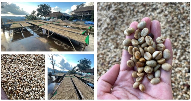
Dr. Nichada Jiannaikun, Beamline Scientist at Synchrotron Light Research Institute (Public Organization) or SLRI, has applied the analysis technique based on synchrotron light to analyze the quality of coffee beans (green beans) or raw coffee beans that have not been roasted. She used Arabica coffee beans that have been processed or dried with different processes such as dry process, wet process, honey process, and the Low temperature and Low humidity (LTLH process) developed by Preeda Coffee Roasting Factory, Lampang Province.
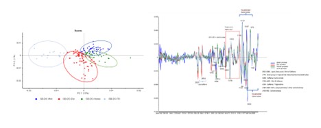
The researchers used synchrotron light combined with Fourier transform infrared spectroscopy which can provide information about the main biochemical components contained in coffee beans including caffeine, fatty esters, Chlorogenic acid and carbohydrate components, and etc. Such substances within coffee beans will be developed into smell and flavor substances when roasted. From the results of the examination (Figure 1), it was found that coffee substances prepared by the dry process had the highest composition of fatty esters. As for the caffeine component, it was found that the amount did not vary in each drying process. When looking at the carbohydrate composition, it was seen that the wash process will have a higher amount of carbohydrates than other processes. As for chlorogenic acid, there is a lot of it in coffee beans that are dried using LTLH Process. This information reveals that the process of preparing coffee substances is different. Therefore, it affects the smell and taste of coffee which is unique to each drying process when roasting coffee substances.
In addition, the research team also conducted a comparative study of the microstructures within coffee bean samples that were dried using four different processes using the X-ray Tomography technique. The results of the analysis (Figure 2) showed that coffee beans prepared by using LTLH Process, developed by Preeda Coffee Roastery, will have a consistent structure within the coffee beans and has the lowest air cavities within the coffee beans, or it has the highest coffee content, compared to coffee substances prepared with other processes.
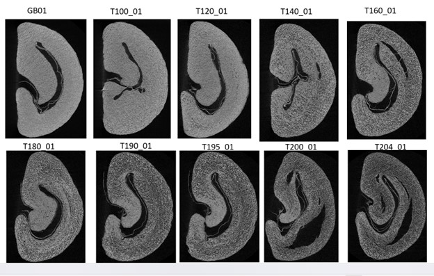
The research team took coffee beans prepared by LTLH Process to study and monitor the microstructural changes within the coffee beans that occur during roasting. The X-ray tomographic imaging technique (Figure 3) compares the internal characteristics of coffee beans (before roasting) with coffee beans at temperatures 100, 120, 140, 160, 190, 195, 200, and 204 degrees Celsius (at the end of roasting). The structure of coffee beans roasted at various temperatures will be useful for coffee developers to find the right roasting style or roasting profile for coffee beans to produce a good smell and flavor.
The structure of coffee beans that have been roasted at various temperatures will be useful for those who are coffee developers in order to find suitable conditions in roasting that will produce coffee with good smell and taste.
The knowledge that we have gained from this study can be recommended or suggested to agriculturists and coffee business entrepreneurs to improve the process related to coffee bean production to gain the most effective quality leading to the elevation of the competitiveness of agriculturists who plant Thai coffee beans. And it will enable Thailand to export Thai coffee beans to worldwide market with high price. This will sustainably result in an increase in coffee beans farmers’ income.
Article by: Dr. Nichada Jiannaikun, Beamline Scientist, Synchrotron Light Research Institute (Public Organization)
Complier: Corporate Communications Section
Translation: International Relations Officer
Figure 1. Utilization of synchrotron light combined with infrared spectroscopy to analyze differences in the biochemical composition of coffee beans prepared in different ways, including the dry process method, shown with a red symbol, the method of washing with water (Wet process) is shown with a blue symbol. Semi-dry, semi-wet method (Honey process) shown with a green symbol, and the drying method at low temperature and humidity (LTLH process) developed by Preeda Coffee Roastery shown with a blue symbol. The left side of the data shows a clear separation into distinct groups. The image on the right shows the different biochemical components within coffee beans prepared from different processes.
Figure 2. It displays three-dimensional photographs of coffee beans using the X-ray Tomography technique which can be used to examine the microstructure or look at the distribution of pores within the coffee beans. The picture below is a comparison of the pores within the coffee beans. The green portion represents the air cavity inside the seed. The black part shows the coffee beans. It can be seen that coffee substances prepared by drying at low temperature and humidity (LTLH) minimizes the formation of cavities in coffee beans.
Figure 3. The image of cut coffee beans obtained by using X-ray Tomography, comparing coffee beans before roasting (coffee substance) and coffee beans at various temperatures. During roasting at 100, 120, 140, 160, 180, 190, 195, 200 and 204 degrees Celsius, it is seen that the pores within the coffee beans increase both in size and quantity, starting at a temperature of 140 degrees Celsius and the cracks that form increase and expand. (Note: The coffee beans photographed are different coffee beans in the roaster.)
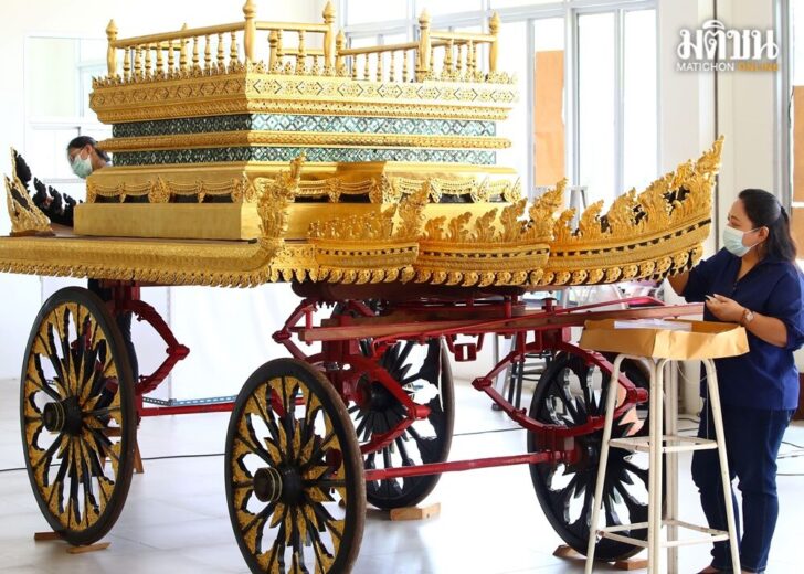
Scientists Restoring Royal Chariot with Synchrotron Light Technology
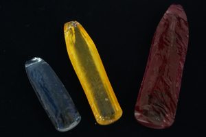
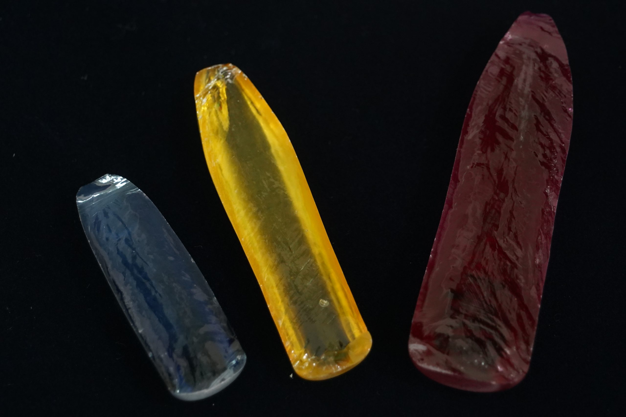
Scientists of Synchrotron Light Research Institute (Public Organization), SLRI, and Ministry of Higher Education, Science, Research and Innovation participated in the restoration of the Royal Chariot which was used in the royal funeral procession or the corpse of the Royal Family, using synchrotron light technology to decipher the formula of the ancient kriab glass and reproduce it to deliver it to Ten Essential Traditional Craftsmanship to renovate and repair before being displayed at the National Museum, Bangkok.
Mrs. Yunee Teeranun, head of the group of gilding and glass decorating technicians and Thai art technicians of Ten Essential Traditional Craftsmanship, Fine Arts Department, stated that “Ten Essential Traditional Craftsmanship is assigned to take care and repair the Royal Chariot นช.539 which is displayed at Bangkok National Museum. And as the time passed by, the Royal Chariot was worn out and the art material that decorated the chariot was gone. Therefore, it needs restoration in order to make the structure strong and the missing pattern will be filled again.”
“However, the glass that is sold in the market has very bright color and it is produced to be used with epoxy glue only. When we restore or repair the artifact in a correct way, we need to choose the original material like Sa-Muk which is the way of using gum tree, coconut shell powder, and burnt banana leaves that are mixed and stirred until it gets sticky, to be used as glue for bonding glass and work piece together. The current glass cannot be used with Sa-Muk and it has different thickness and thinness from the original glass.”
“Therefore, Ten Essential Traditional Craftsmanship has requested SLRI to produce glass that is similar in color and thickness to the original glass on the Royal Chariot. And when we use the produced glass with Sa-Muk, it works well together. After we complete the restoration, we will put on to display at Bangkok National Museum.” – Mrs. Yunee Teeranun.
Dr. Wantana Klysubun, Assistant Director for Academic and Research Affairs, SLRI, said, “This work is an extension of research on the study of kriab glass from Wat Phra Sri Rattana Satsadaram (Wat Phra Kaew), which was conducted for many years at that time until it was successful in gaining knowledge and the formula of the glass which can help produce the ancient glass in four colors. That is the reason why we got contacted from Ten Essential Traditional Craftsmanship that they have the project of restoring the Royal Chariot. And it is a conservation restoration which means they will not remove cracked glass. They will preserve the old ones; however, the missing ones will be replaced with newly produced glass that has the same color and thinness compared to the original ones. The newly produced glass can be used and endure with gum tree which is a natural glue that is the ancient technique which is used with wood. The glass that SLRI produced has got all the characteristics. We can adjust the color, come up with new glass formula; that is why we got this assignment. We had been working on it for around one year to adjust the color of the glass formula since the green and blue color of the Royal Chariot are slightly different from crystal glass from Wat Phra Sri Rattana Satsadaram (Wat Phra Kaew). Therefore, the colors are adjusted in order to create the new ones to be similar to the original ones, as you can see.”
“SLRI is solely responsible for producing glass. There are many colors that were delivered to us. There are green jewel beetle color that is slightly bluish and off-white-yellow. We created a new specific formula which has blue, red, and orange. For the red-orange color, it is very interesting. Because the technician said it was white glass before. But after the restoration, another color was found hidden under the white glass. They sent it to us so we could see that it was reddish orange. This red-orange color is very challenging. Since it is not the color found at Wat Phra Kaew. It is a new ancient color which we have never studied before. Therefore, we expand the knowledge on the ancient red color and antique yellow by mixing them together, thus, this new glass color can be produced. And then we send to the Ten Essential Traditional Craftsmanship to use.
“Using Synchrotron is like starting closer to the goal. It acts as a shortcut, allowing us to achieve results faster. Because if we needed to begin from the basic formula, it would be so difficult, especially ancient glass formulas. The color composition is very complex. It is not just only one element which can create the color; however, it takes 2 – 3 elements. Thus, we need to adjust to build the balance to create the color’s uniqueness for the ancient fine art. Notwithstanding, the expansion to the level of pilot plant requires the collaboration with private sectors who are interested in further developing the possibility of commercial production.” Dr. Wantana Klysubun concluded.
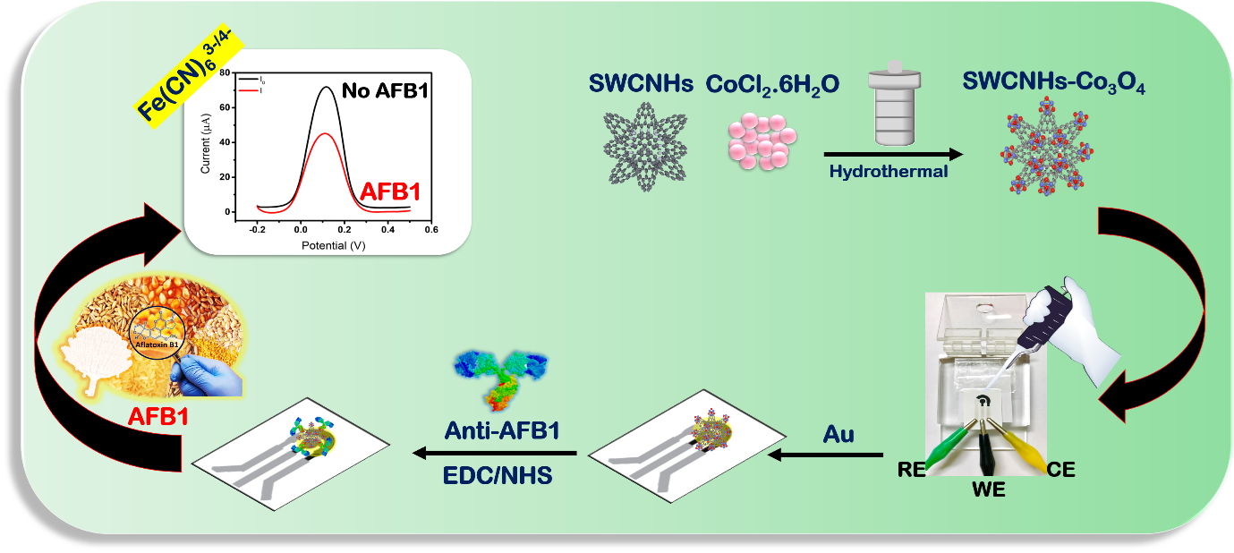
Figure 1 Preparation procedures of the proposed AFB1 immunosensor for AFB1 detection
Mycotoxins are widely found in daily products such as peanuts, beans, corn, wheat and rice. Aflatoxin B1 (AFB1) is one of the most toxic which may affect a serious harm to human and animals, thus causing both acute and chronic diseases.
Therefore, a novel sensitive and selective platform for the specific determination of aflatoxin B1 (AFB1) was developed by Associate Prof. Dr. Anchalee Samphao (Department of Chemistry, Faculty of Science, Ubon Ratchathani University).
Anti-AFB1 antibody was immobilized on single-walled carbon nanohorns decorated by cobalt oxide composite and gold nanoparticles as shown in Figure 1. Thereafter, selectivity of the proposed sensor was attributed to the specific binding property of anti-aflatoxin B1 antibody.
X-ray crystal structure (PDB entry 5B3N) was used as a structural template for a single-chain fragment variable (scFv) of monoclonal antibody against aflatoxins. The specific interaction of the proposed immunosensor to AFB1 was clarified using homology modelling combined with molecular docking. In the presence of AFB1, the current signal of the modified electrode became lower which involved specific antibody-antigen binding (Figure 1), including hydrophobic, hydrogen bonding and pi-pi stack interactions.
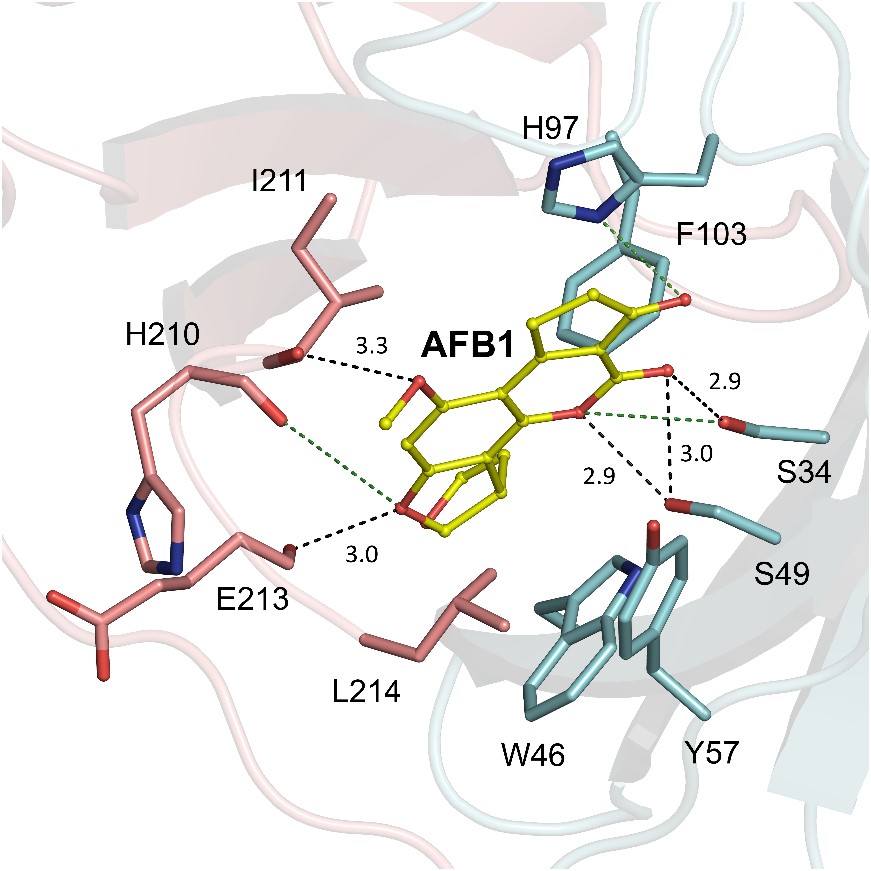
Close inspection of the predicted structure based on 5B3N revealed that heavy chain (VH) and light chain (VL) contributed to the aflatoxin binding site as shown in Figure 2. It is found that docked molecule of AFB1 interacts at the surface between the variable domains of both VH and VL (Figure 2), thus contributing to the antigen-binding site.
Figure 2 The interactions between antibody and aflatoxin B1. The model structure based on 5B3N is shown in cartoon representation whereas heavy chain (VH) is shown in cyan and light chain (VL) is shown in salmon. The labelled amino acid residues are shown as stick models with carbon atoms in cyan (VH) and salmon (VL), nitrogen atoms in blue and oxygen atoms in red. Aflatoxin B1 is shown as a ball-and-stick model with carbon atoms in yellow and oxygen atoms in red. Black dash lines represent hydrogen bonds formed with their distances given in Å. Electrostatic interactions are represented by green dash lines.
Among four kinds of aflatoxin, it showed that AFB1 has the highest affinity towards antibody, thus considering the binding energy of AFB1.
The application of the proposed immunosensor in real samples, including peanut, certified reference material of peanut sample (labelled 206 µg kg-1 AFB1), corn, and chicken feed was investigated. This present work shows an excellent performance for AFB1 detection which could be applied for food quality control as well as modified to detect other mycotoxins.
Re-written by Chomphunuch Songsiriritthigul (Beamline Scientist, SLRI)
References
Benkerroum, N. Chronic and acute toxicities of aflatoxins: Mechanisms of action, Int. J. Environ. Res. 17 (2020) 423.
Damphathik, C., Songsiriritthigul, C., Lerdsri, J., Jakmunee, J., Wongnongwa, Y., Jungsuttiwong, S., Ortner, A., Kalcher, K., Samphao, A. A novel immunosensor based on cobalt oxide nanocomposite modified single walled carbon nanohorns for the selective detection of aflatoxin B1. Talanta. (2023). DOI: 10.1016/j.talanta.2023.124472.
Moretti, A., Logrieco, A.F. & Susca, A. Mycotoxins: An Underhand Food Problem, in: A. Moretti, A. Susca (Eds.) Mycotoxigenic Fungi: Methods and Protocols, Springer, New York, New York, NY, 2017, pp. 3-12.
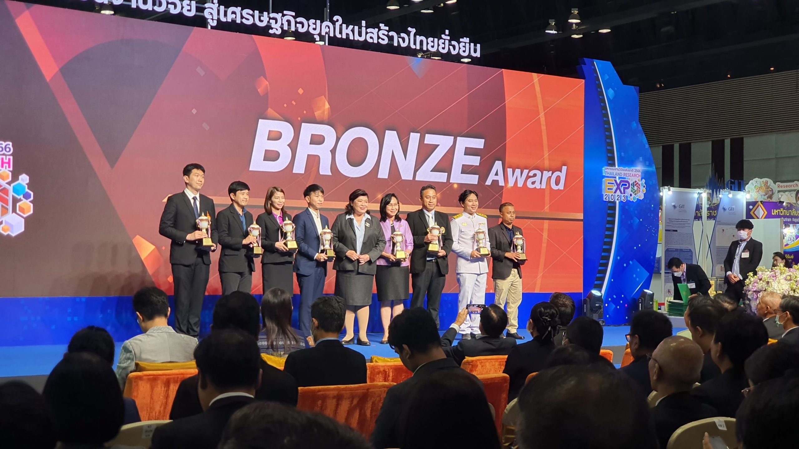
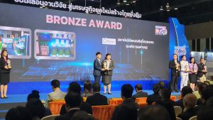
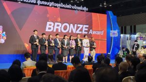
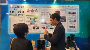
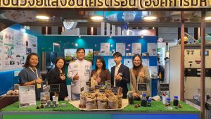
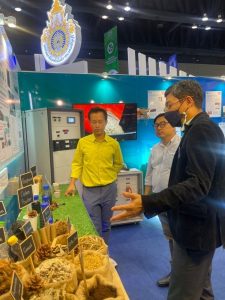
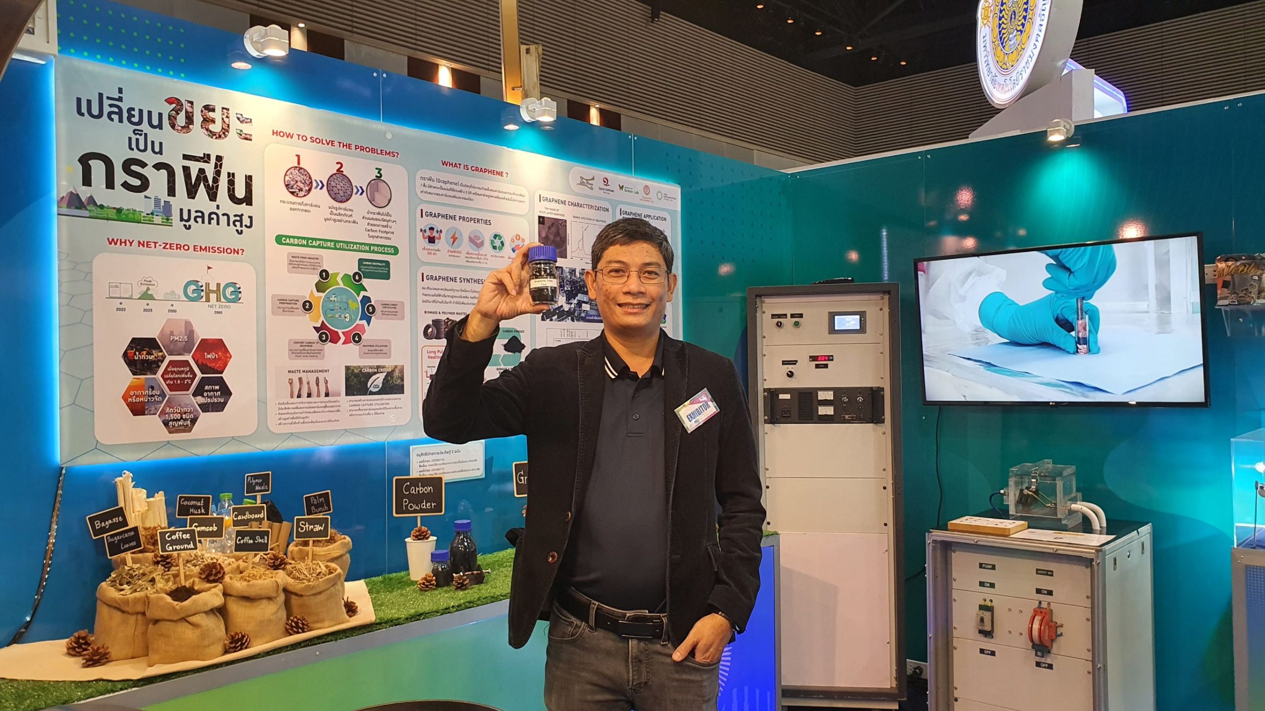
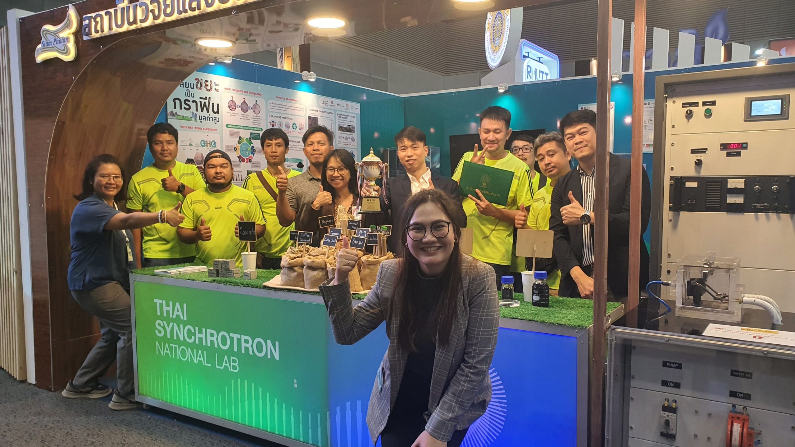
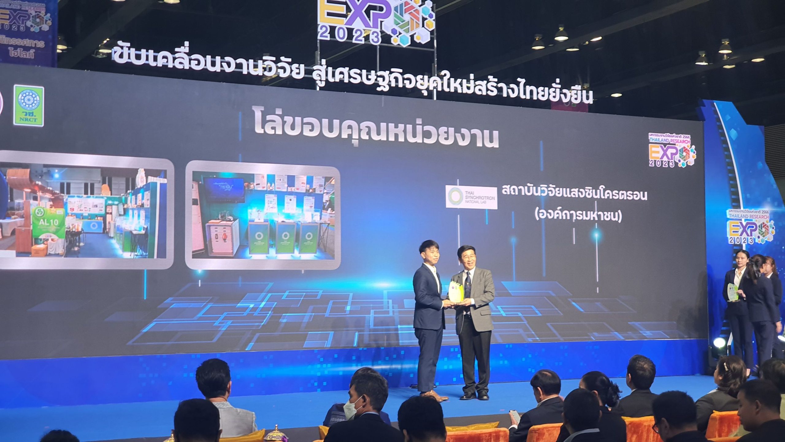
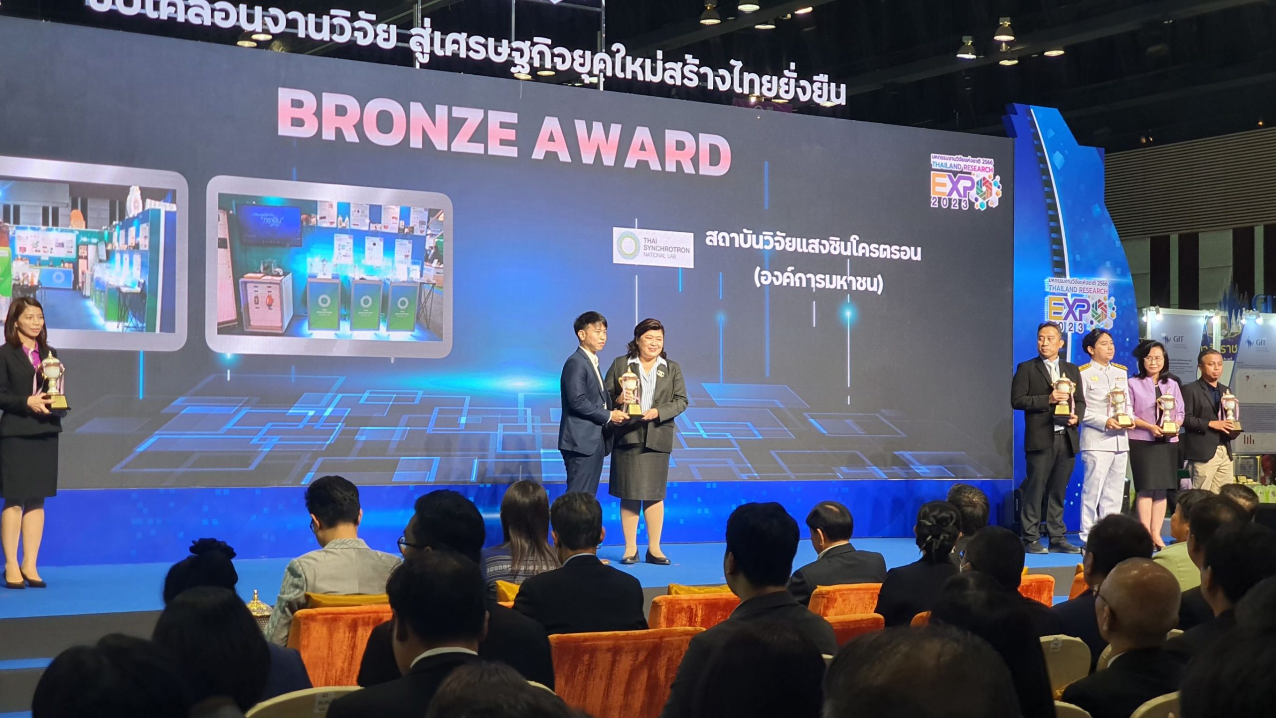
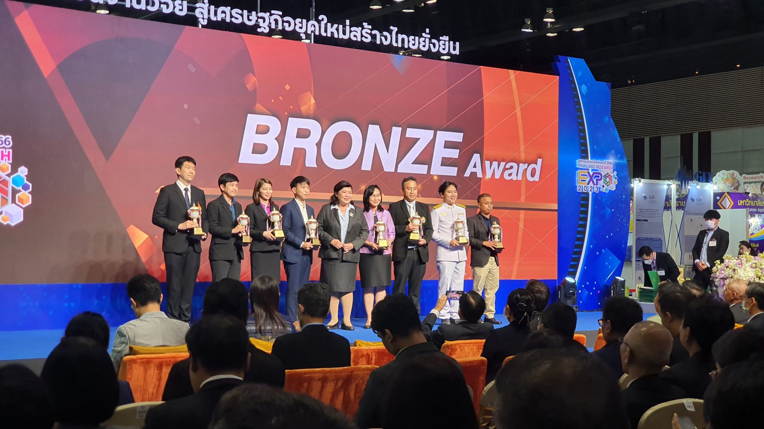
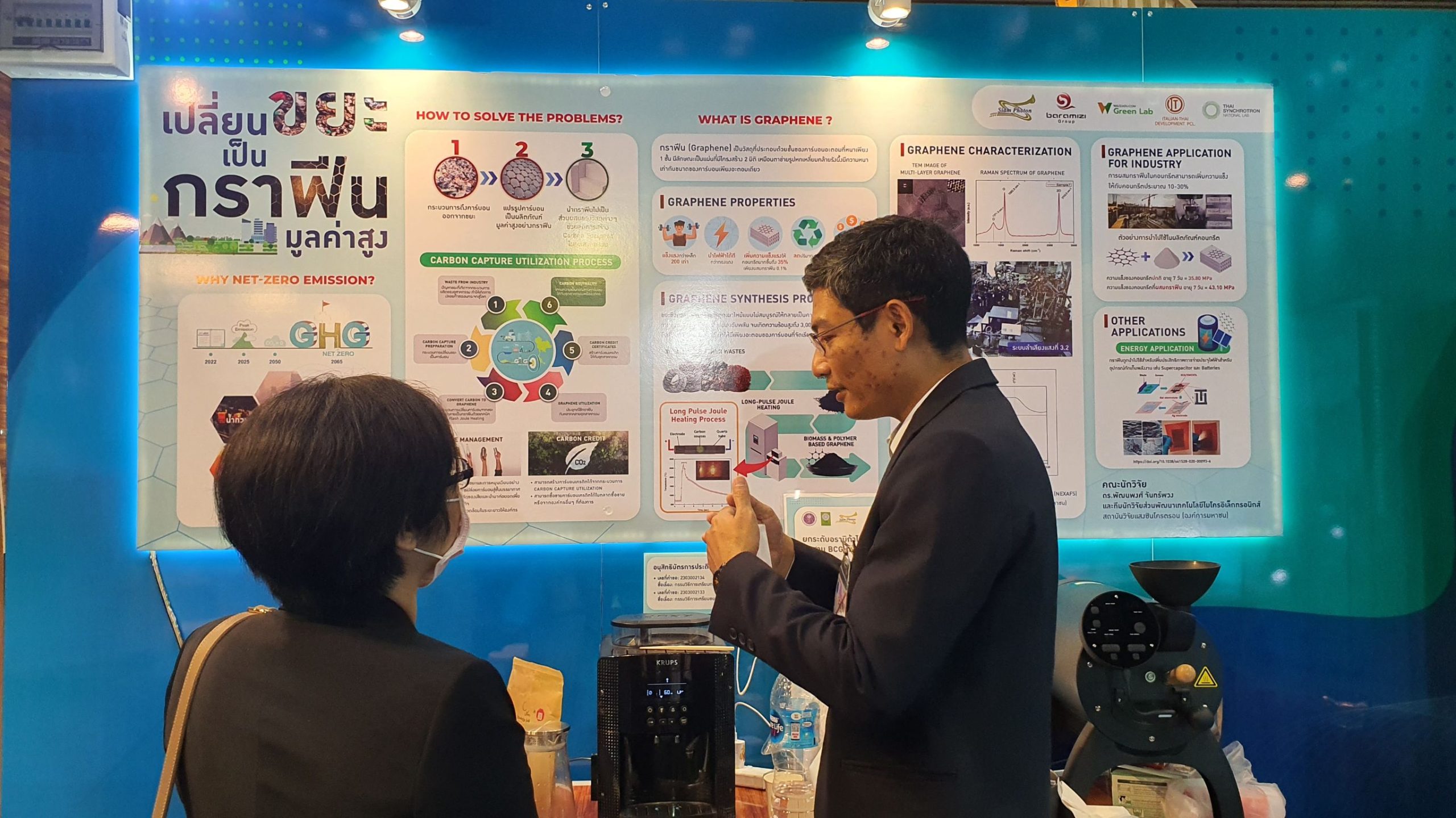
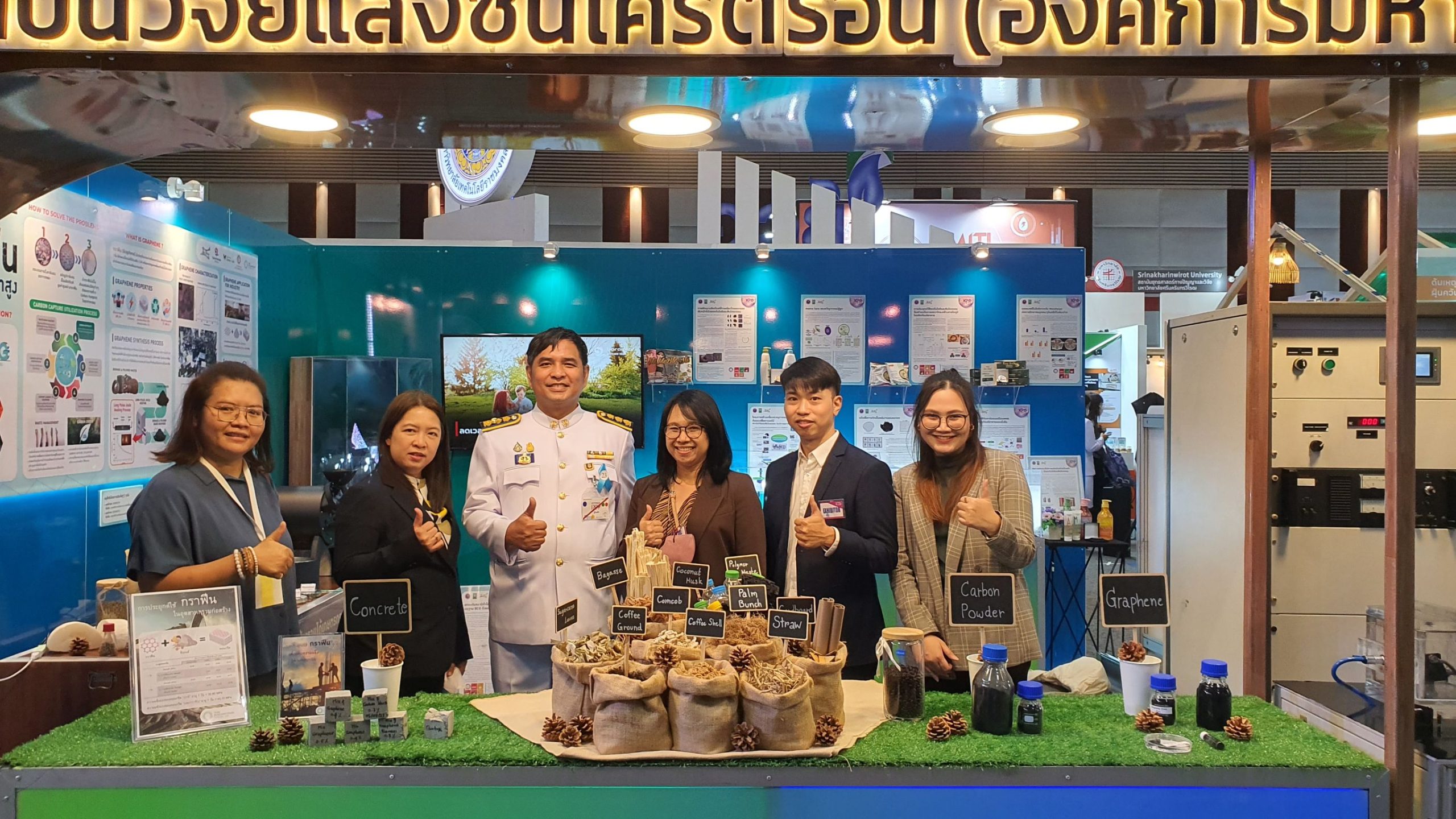
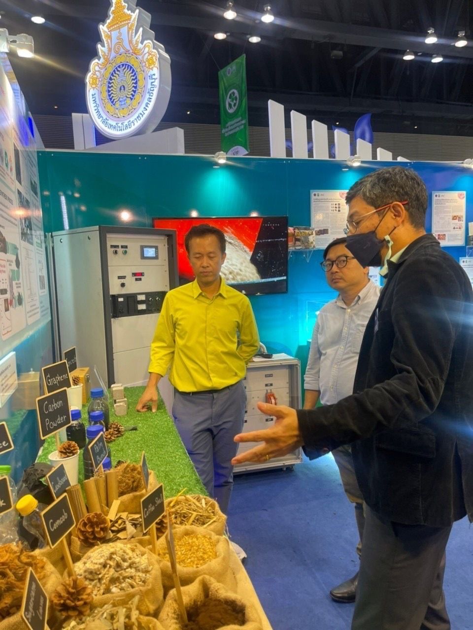
The research project of transforming waste into “graphene” done by Synchrotron Light Research Institute (Public Organization) or SLRI, Ministry of Higher Education, Science, Research and Innovation (MHESI) is achieved with the Bronze Award at the National Research Exhibition 2023. The project aims to transform waste into future valuable materials.
Dr. Sarayut Tunmee, Chief, Organization Strategy Division of SLRI, stated, “the work of turning waste into high-value graphene is a significant research achievement. Dr. Pattanaphong Janphuang, Chief, Technical and Engineering Development Division of SLRI, wins the Bronze Award for this project at the National Research Exhibition 2023 organized by the National Research Council of Thailand (NRCT) on 7th – 11th August 2023, at the Centara Grand and Bangkok Convention Centre at Centralworld, Bangkok.”
Dr. Tunmee said, “The project involves converting waste into graphene by using rapid thermal process on selected waste rich in hydrocarbons as main components such as saline bottles, plastic bottles, car tires, coconut husks, and corn cobs. This project helps reducing biomass waste incineration, handling waste treatment, and recycling carbons for future use. For instance, graphene can be mixed into concrete to enhance its strength which supports achieving the Net Zero emissions goal to reduce global warming. This project also contributes to turning waste into high-value materials of the future.”
“This research is among many research based on the Bio-Circular-Green Economy (BCG) concept presented by SLRI at the exhibition this year. Other research examples include the quality improvement of Thai Arabica coffee beans using synchrotron light, development of glass as new and environmentally friendly battery material, and development of diamond-like carbon as environmentally friendly packaging,” added Dr. Tunmee.
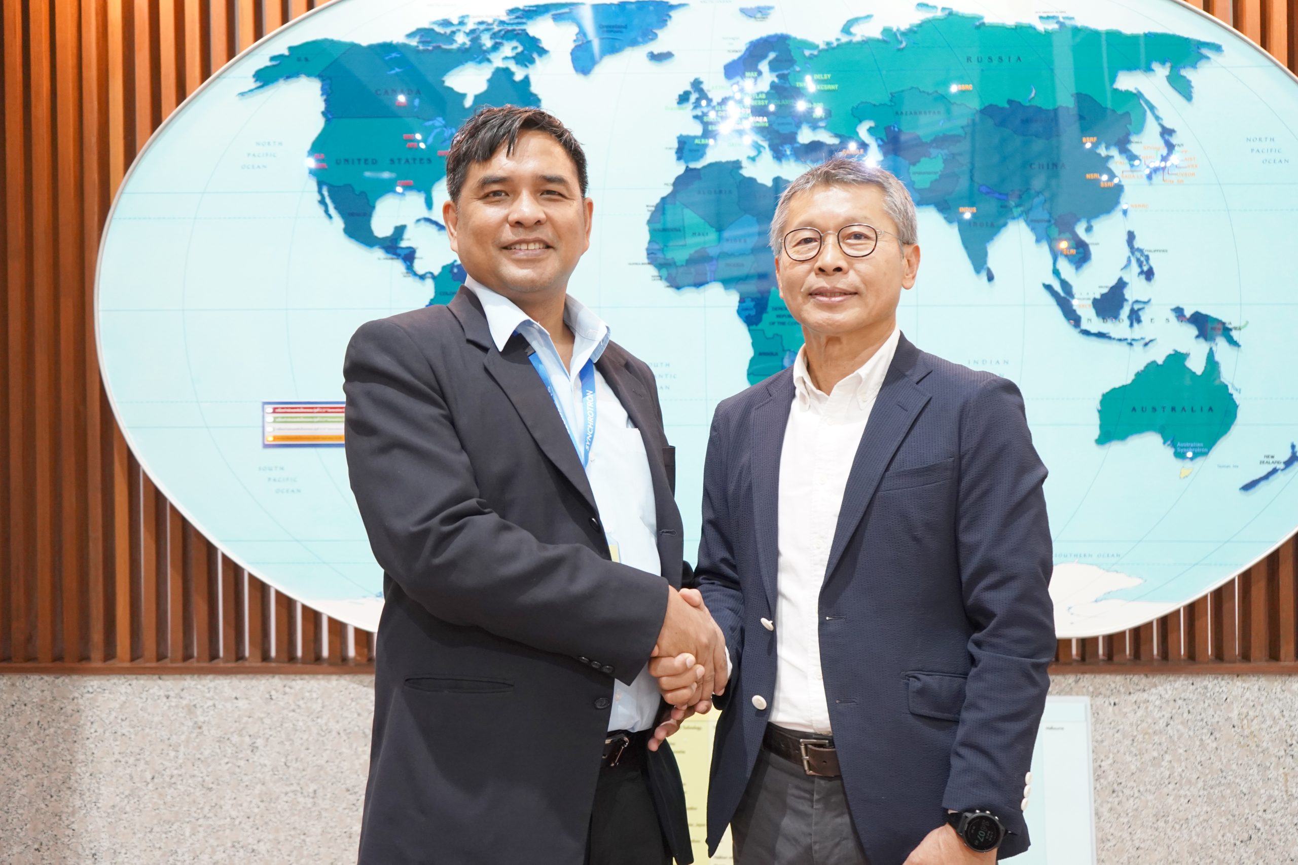
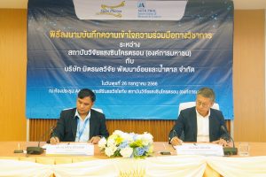
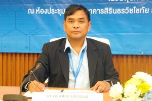
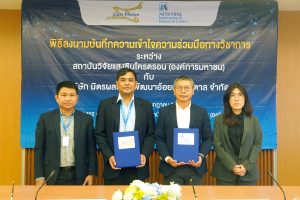
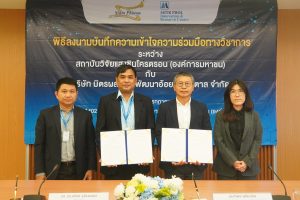
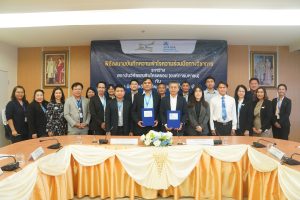
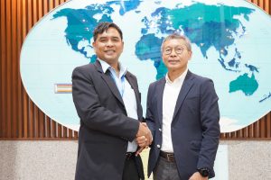
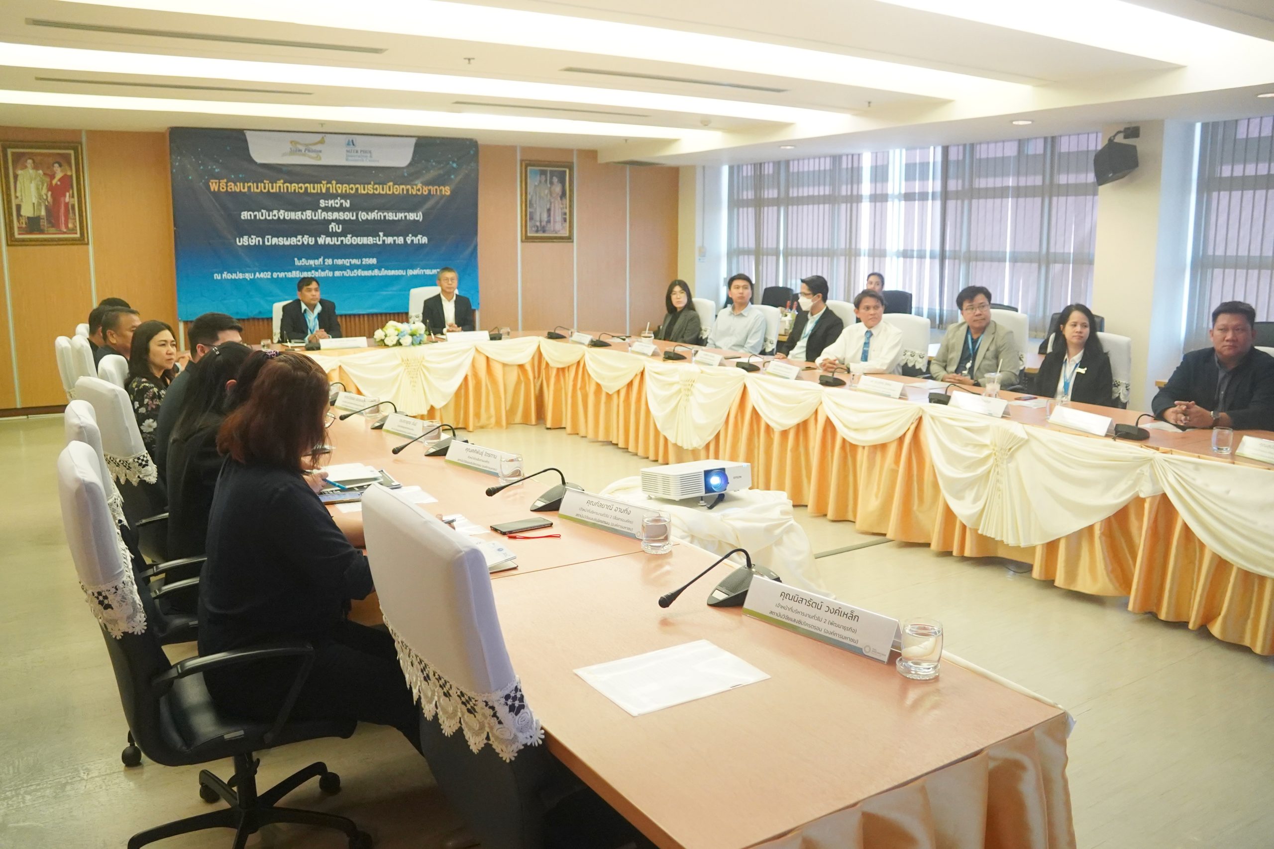
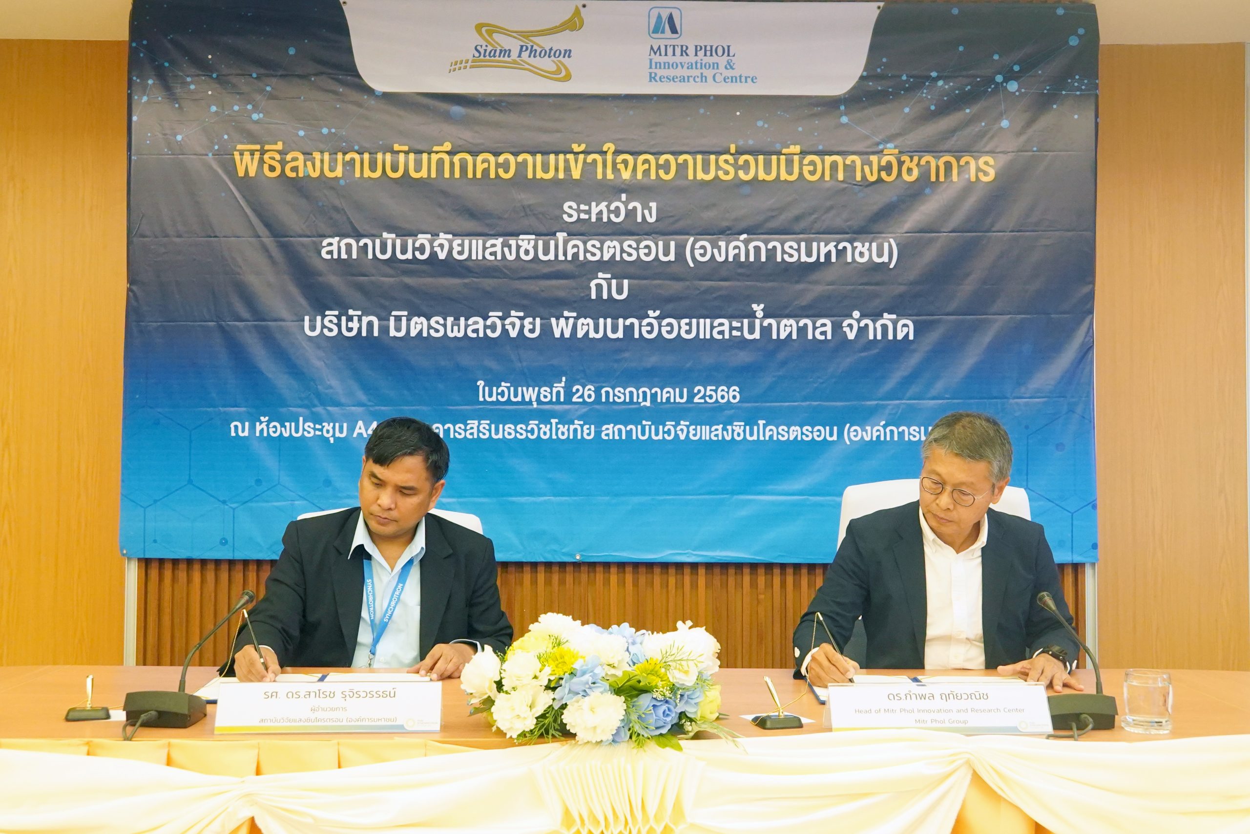
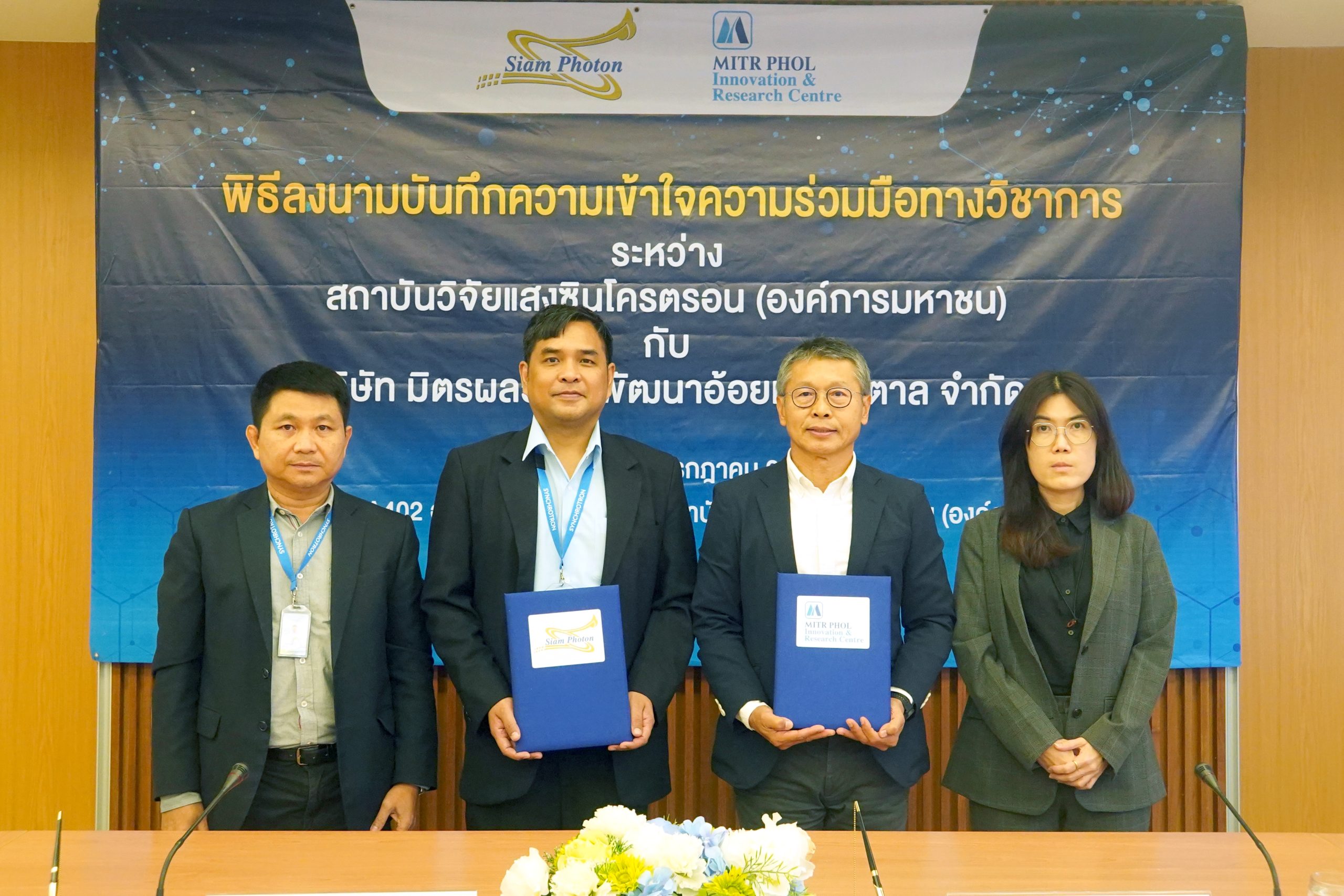
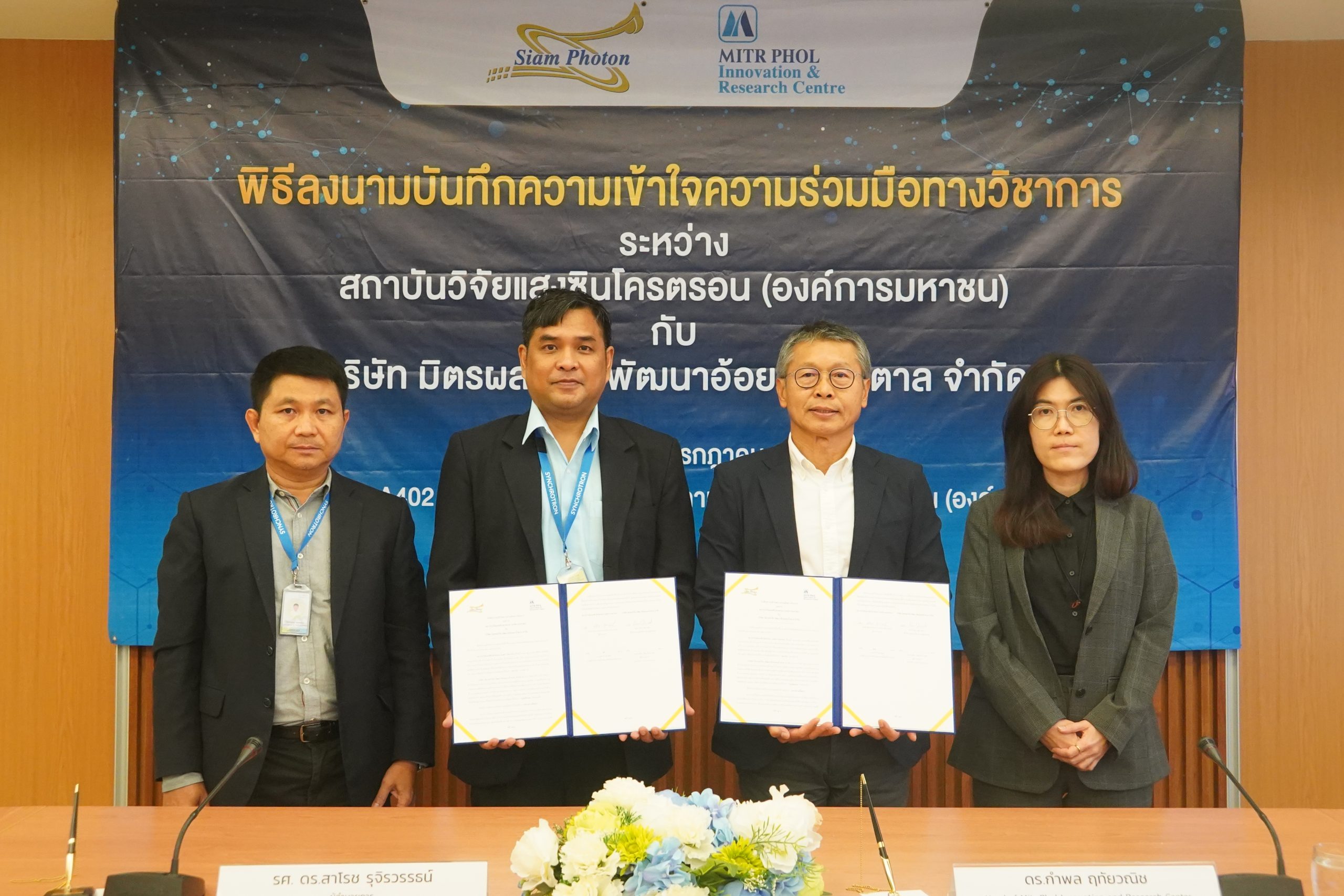
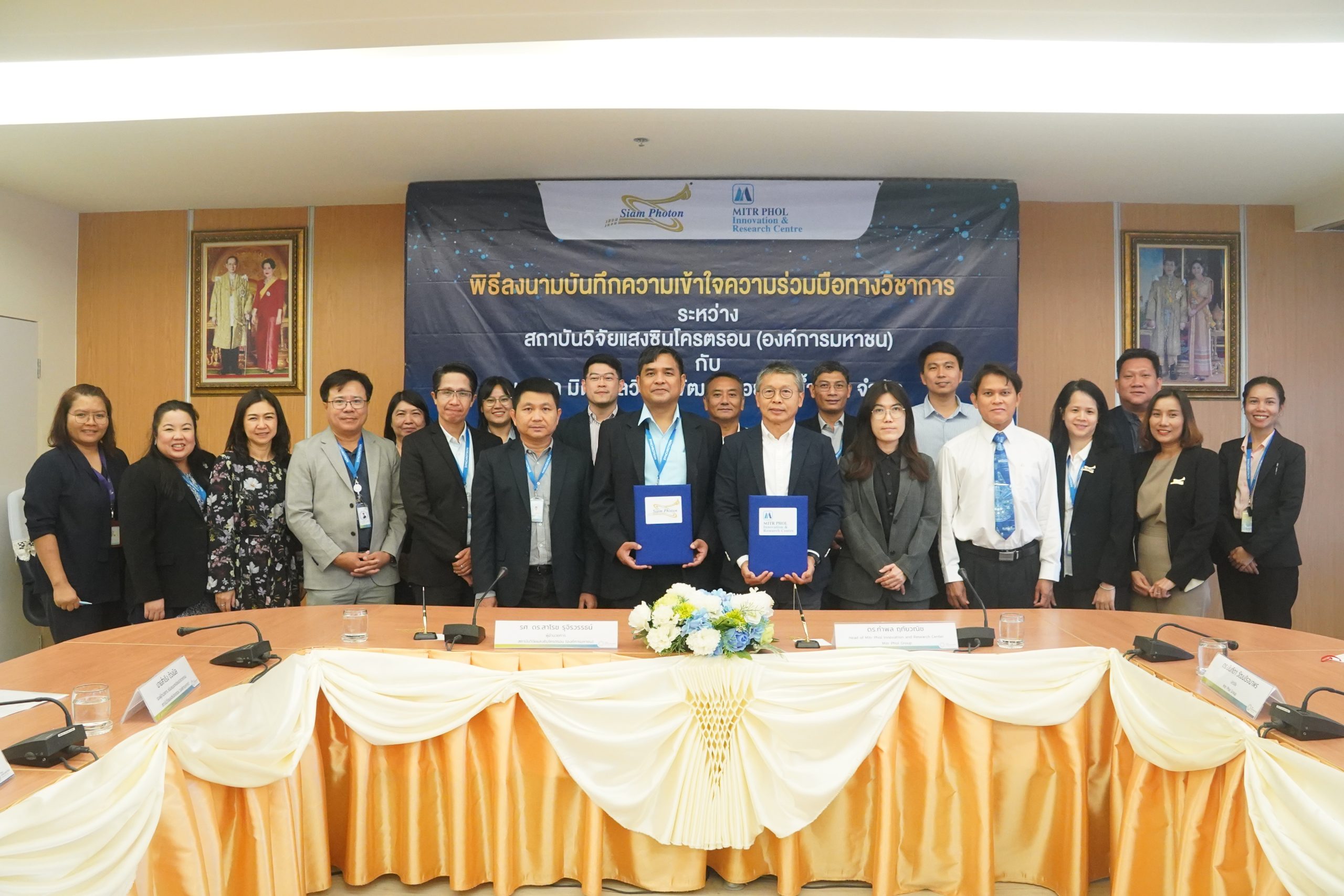
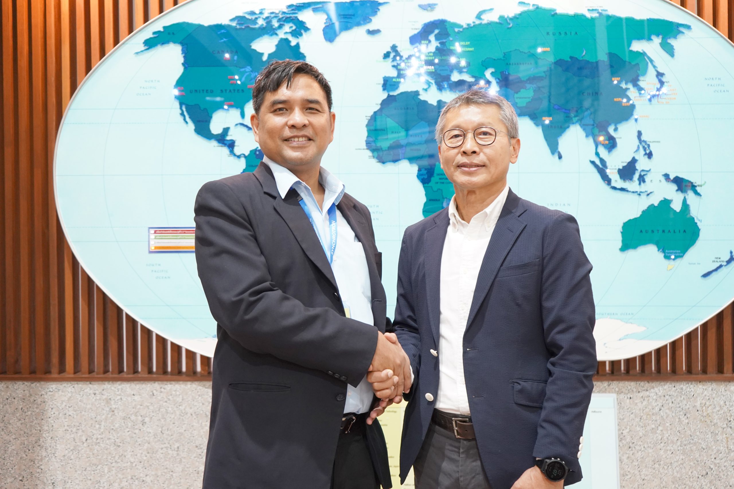
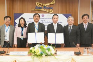
Dr. Kampon Ruethaivanich, Consultant of Mitr Phol Innovation & Research Center, Mitr Phol Innovation & Research Center Co., Ltd. and Assoc. Prof. Dr. Saroj Rujirawat, Director of Synchrotron Light Research Institute (Public Organization) or SLRI, Ministry of Higher Education, Science, Research and Innovation (MHESI) signed the Memorandum of Understanding (MoU) on Wednesday 26th, July 2023 at Sirindhornwitchothai building, SLRI, Nakhon Ratchasima in order for the two organizations to collaborate in academics, and product research and development based on the Net Zero approach for lower carbon release.
Dr. Ruethaivanich stated, “the company and SLRI conduct ongoing research on producing high-value products out of raw materials or waste to promote the company’s growth and increase competitive advantage. The company also aims to work with SLRI on the current agricultural research including production related to sugar and sugarcane as well as production of biomass electricity, ethanol, healthy products, pet food products, and extracts.”
“Mitr Phol Innovation & Research Center Co., Ltd. highly recognizes the importance of environmental policy and Net Zero emission is taken into account as part of the company’s strategy since our products are sold worldwide and carbon emission issue is raised in awareness among our customers. Our environmental policy will therefore affect customers’ consideration and our competitiveness later on. The company conducts research on carbon emission reduction, database of total reduced carbon, and environmental friendliness of our products. We are pretty sure that SLRI will be our active partner in Net Zero policy”, said Dr. Ruethaivanich.
Assoc. Prof. Dr. Rujirawat said, “This MOU includes working together on synchrotron-related research, development, technology, equipment, and tools. The MOU signing will lead to collaboration in research analysis, analyzing techniques, and laboratory tools, from laboratory to prototype and industrial scales. Mitr Phol Innovation & Research Center Co., Ltd. and SLRI will also support each other in academics, trainings, human resources development, techniques and engineering.”
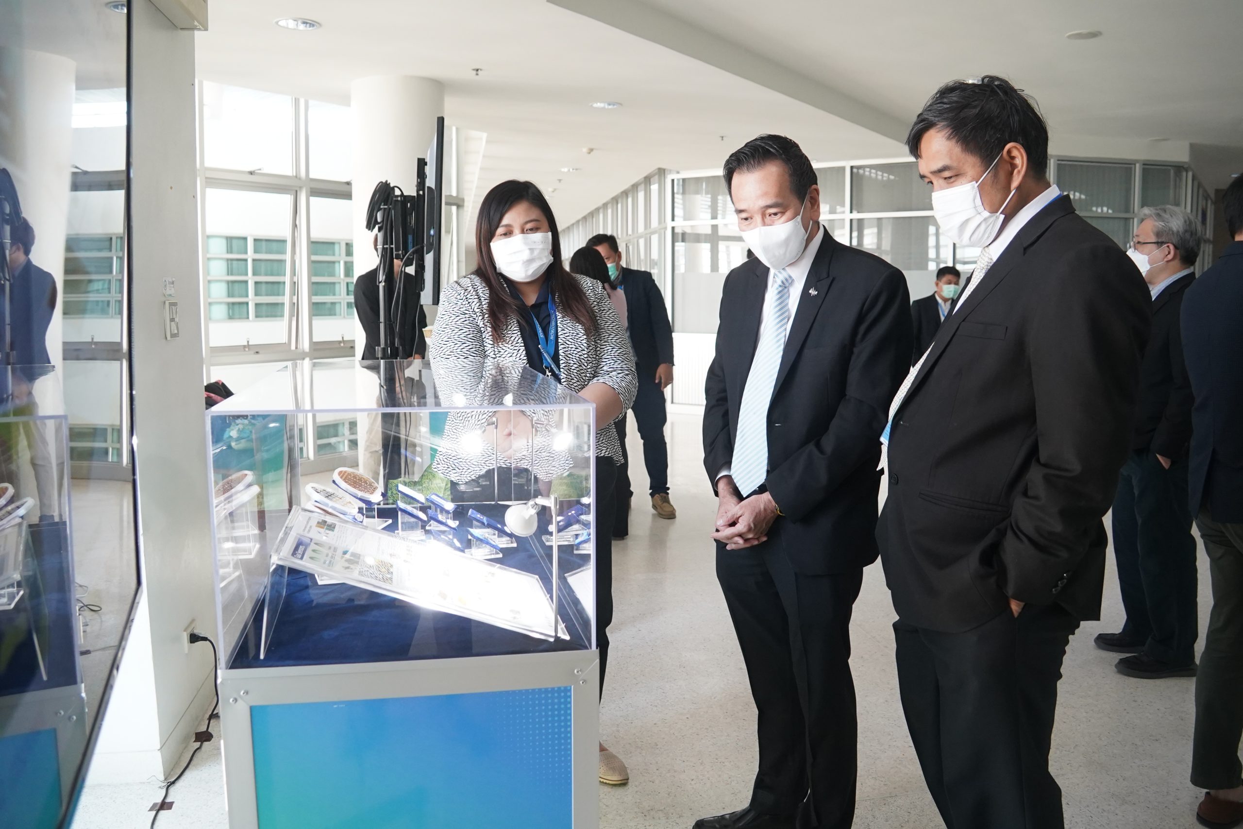
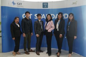
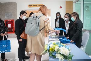
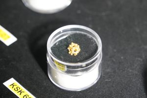
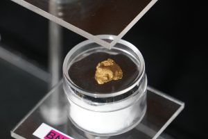
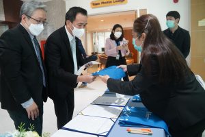
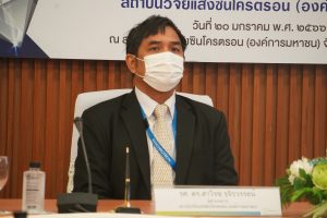
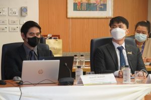
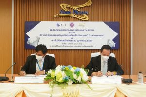
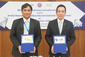
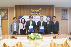
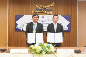
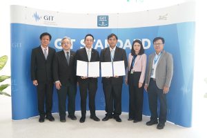
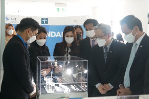
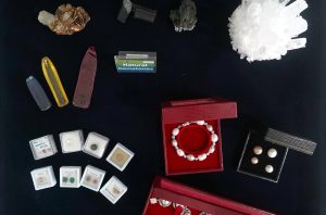
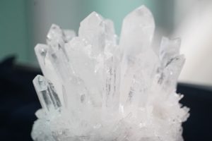
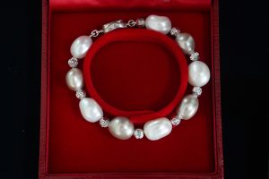
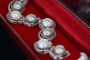
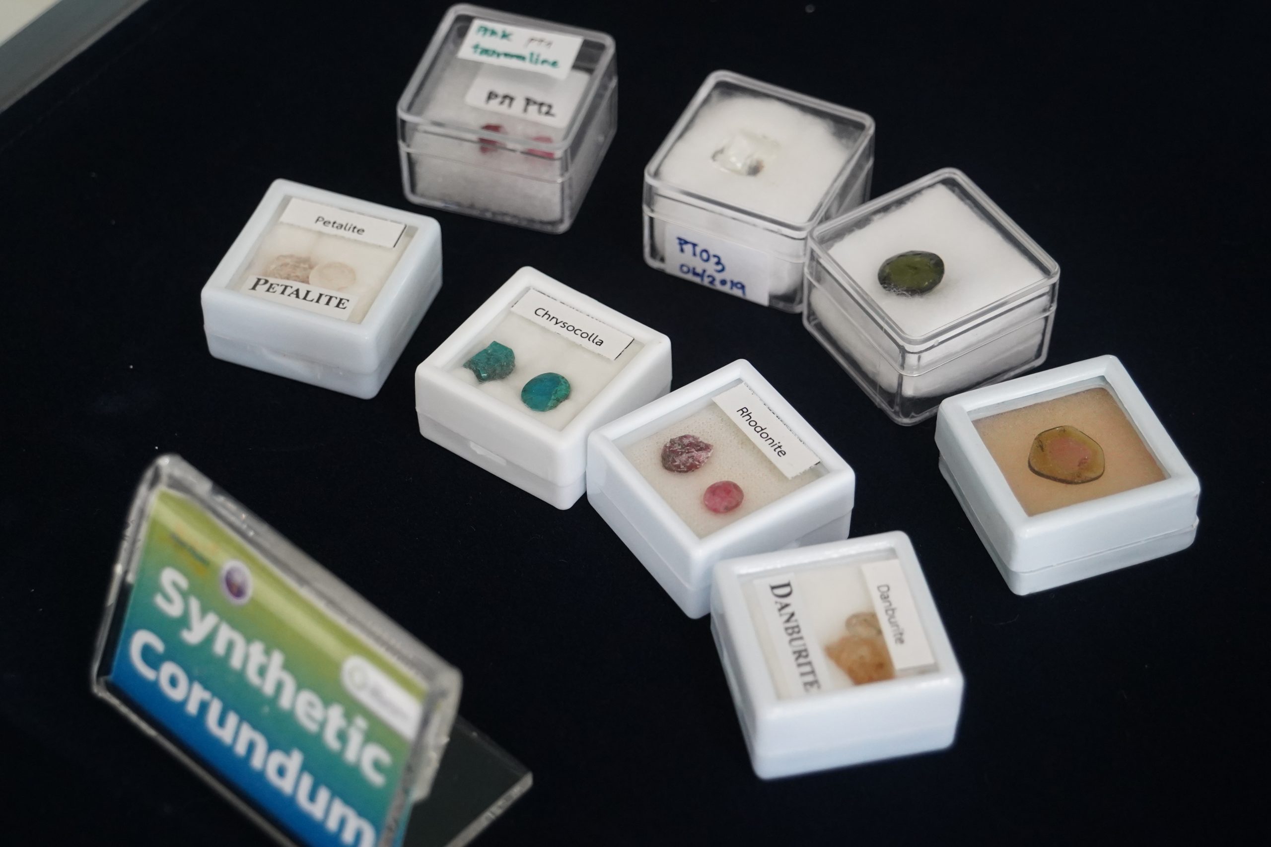
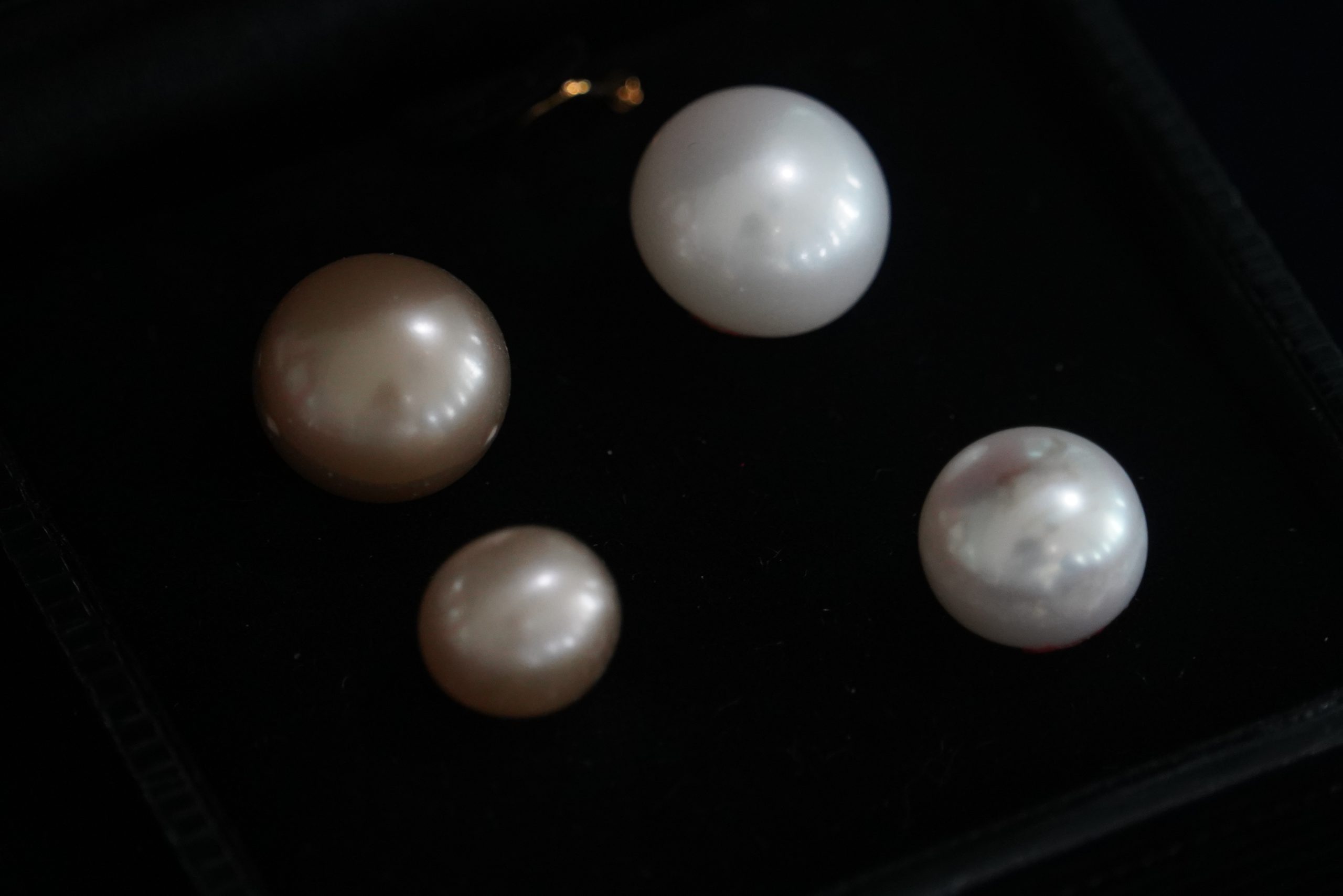
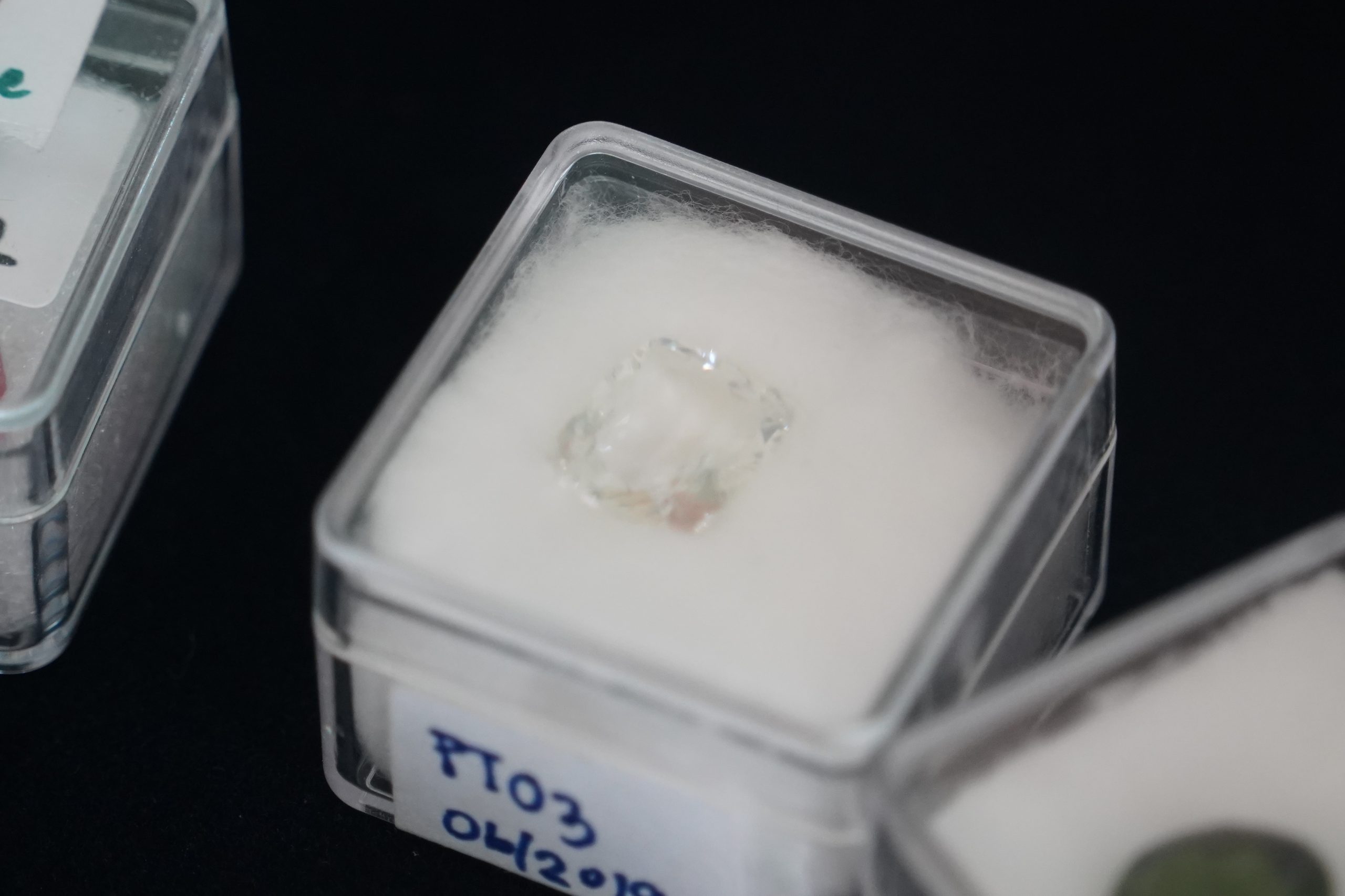
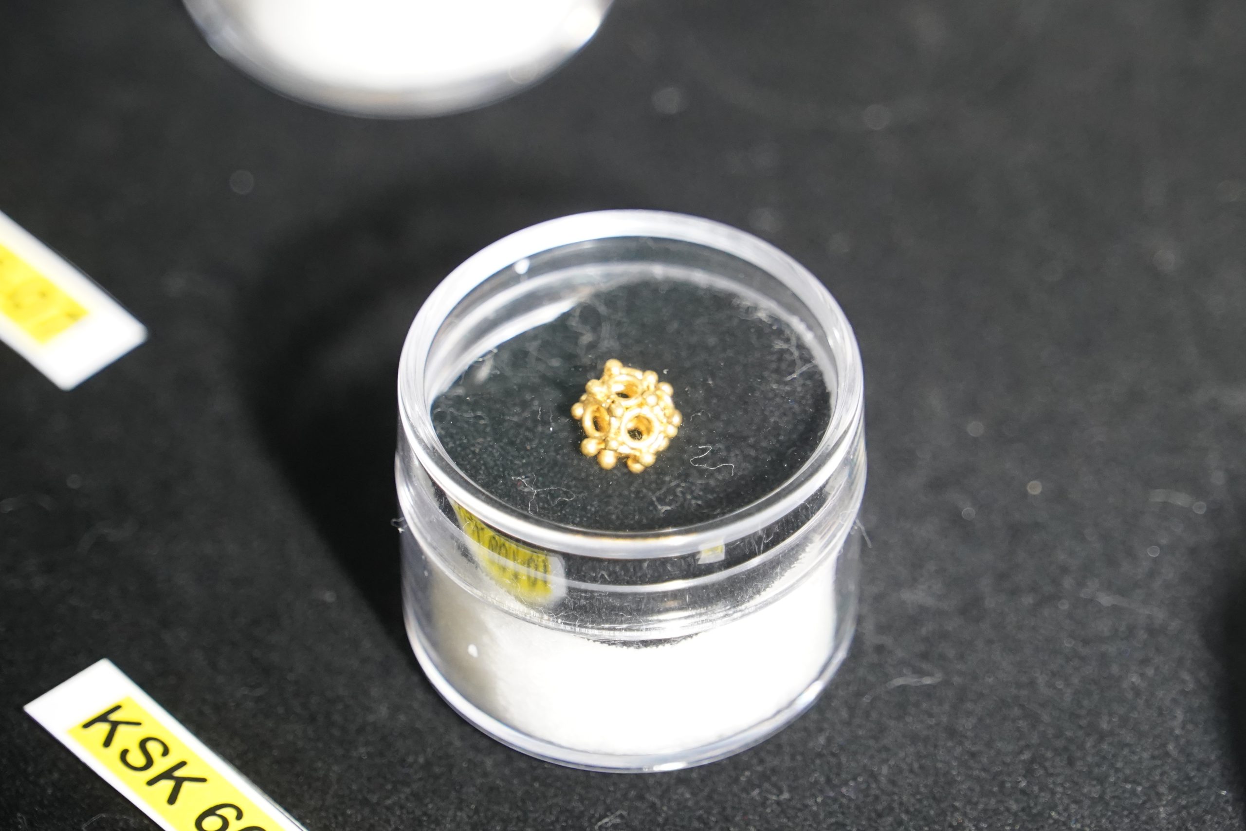
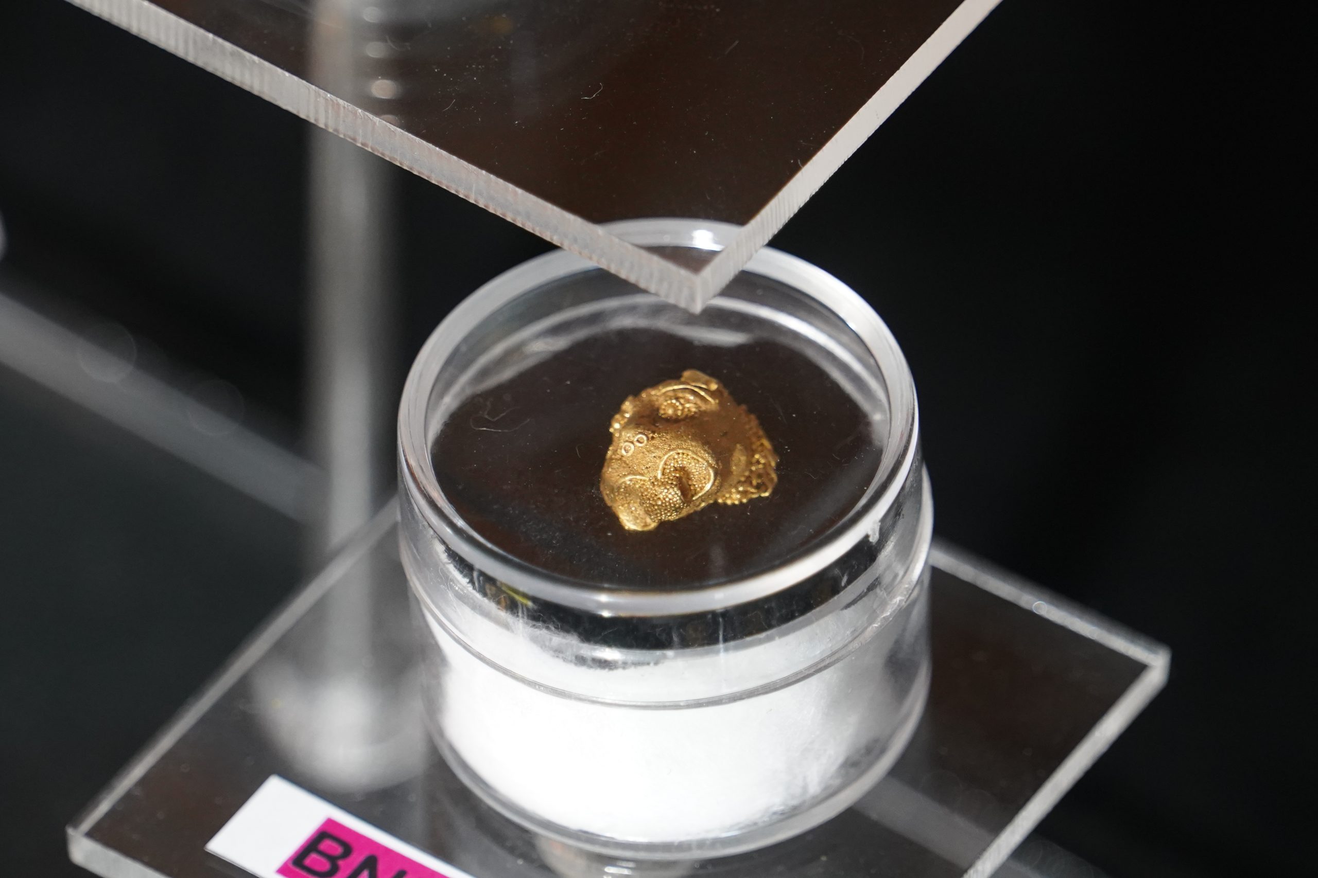
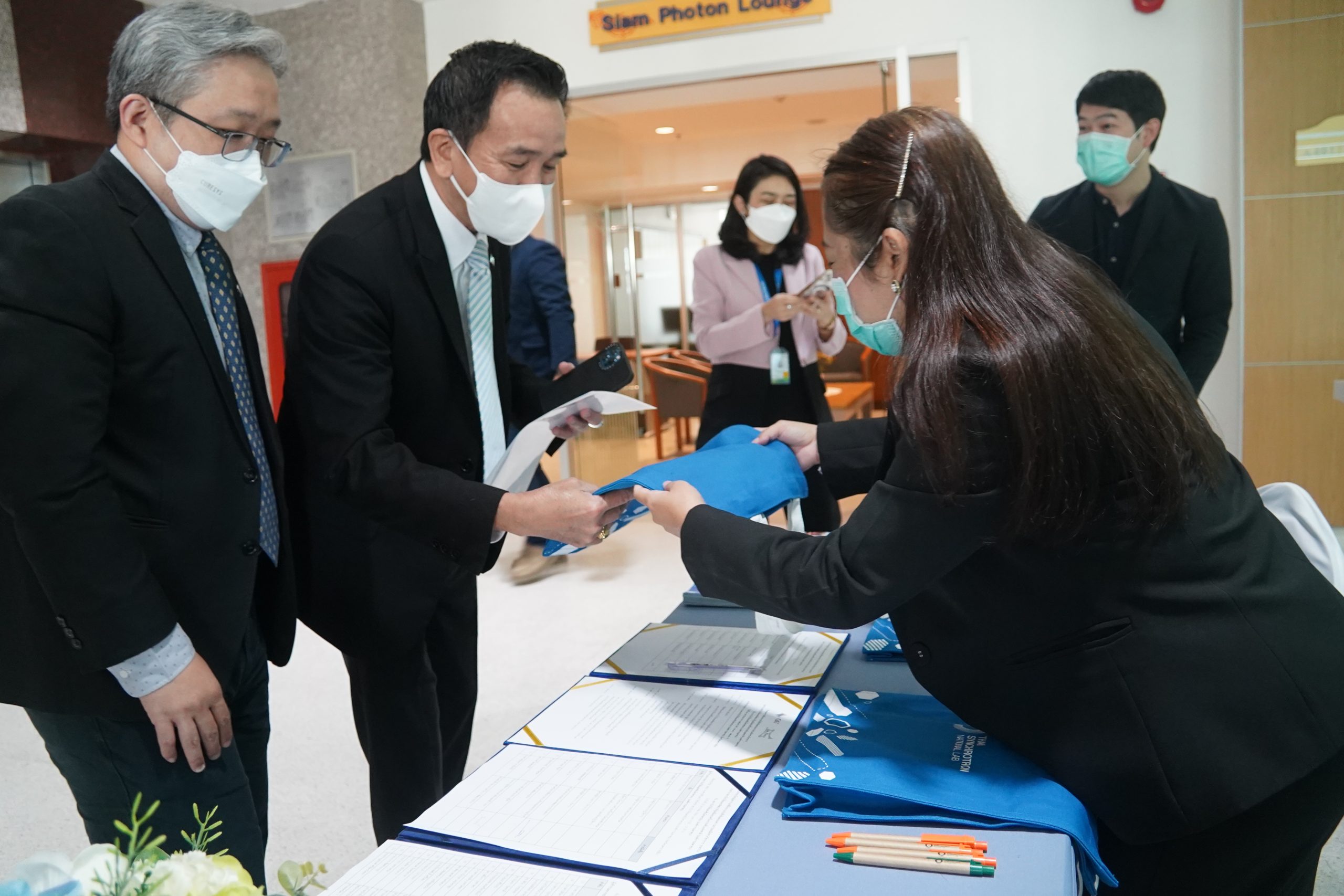
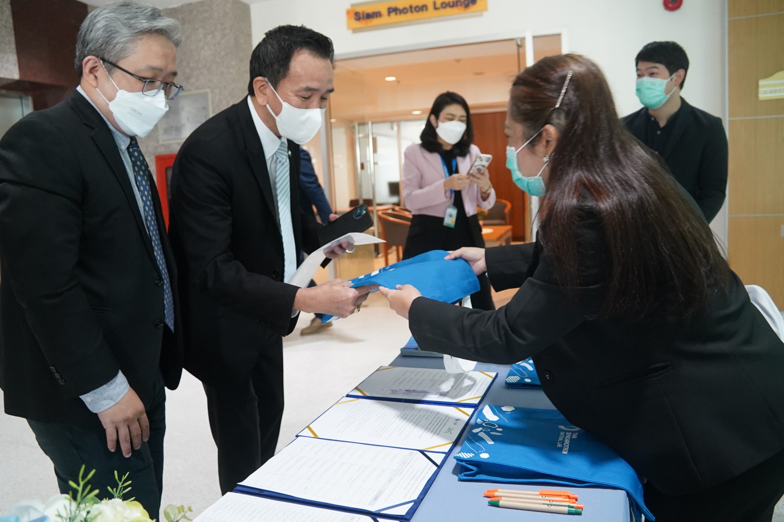
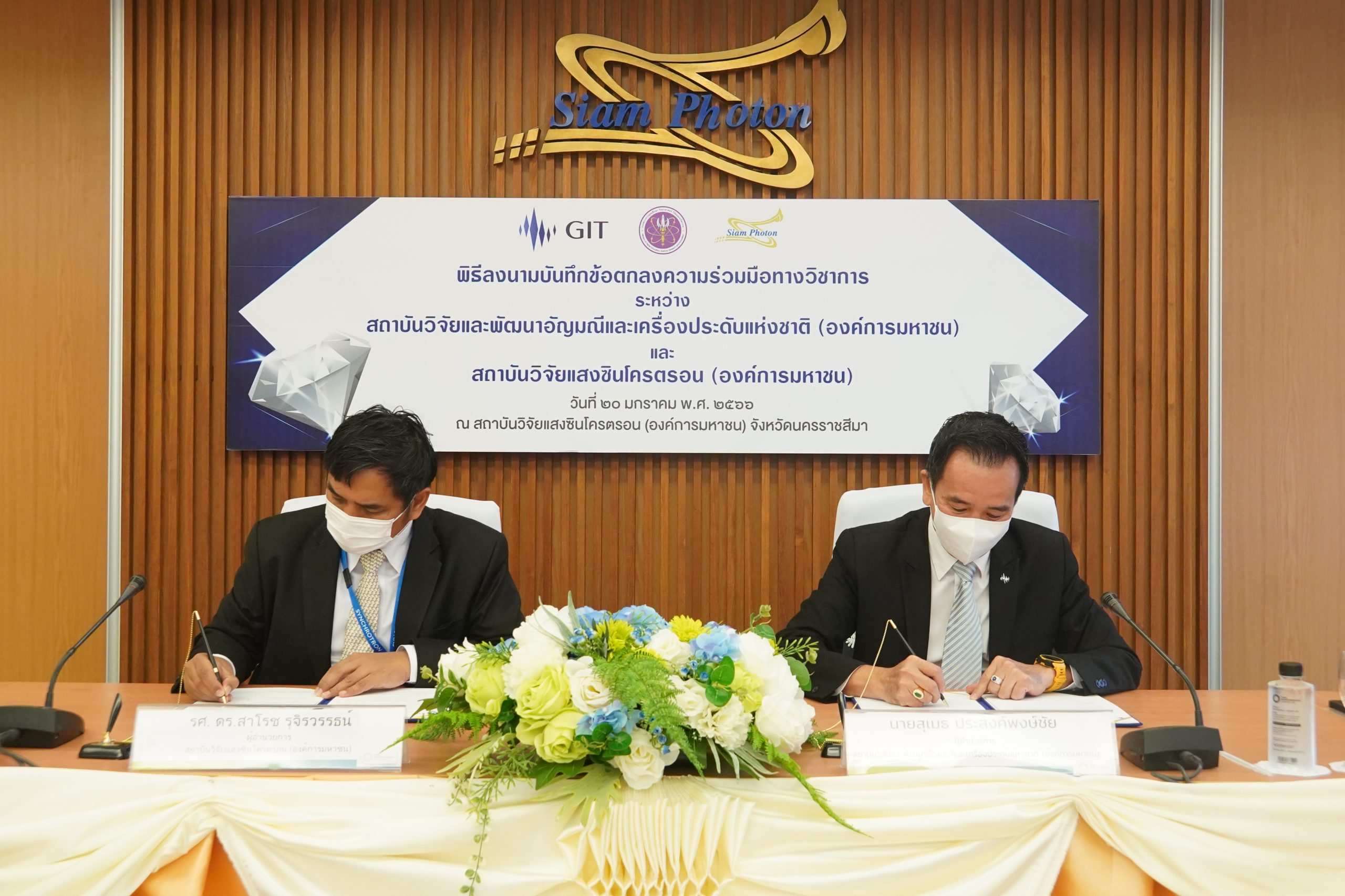
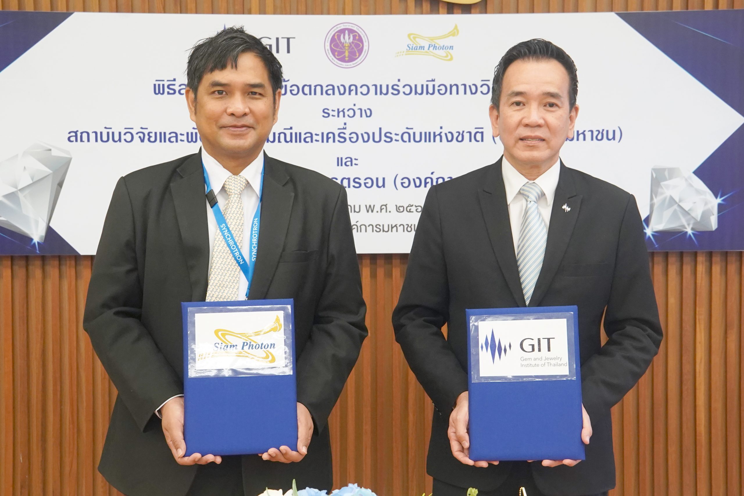
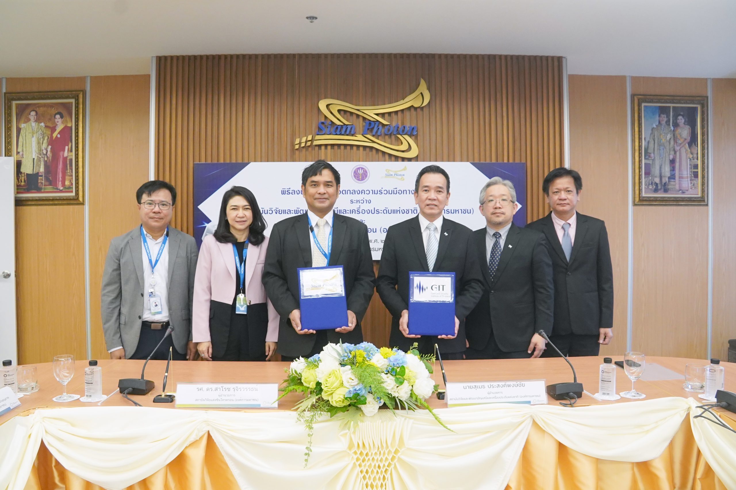
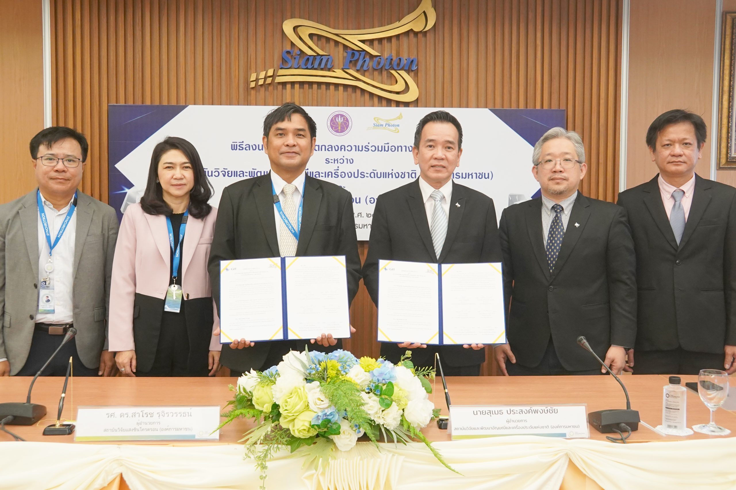
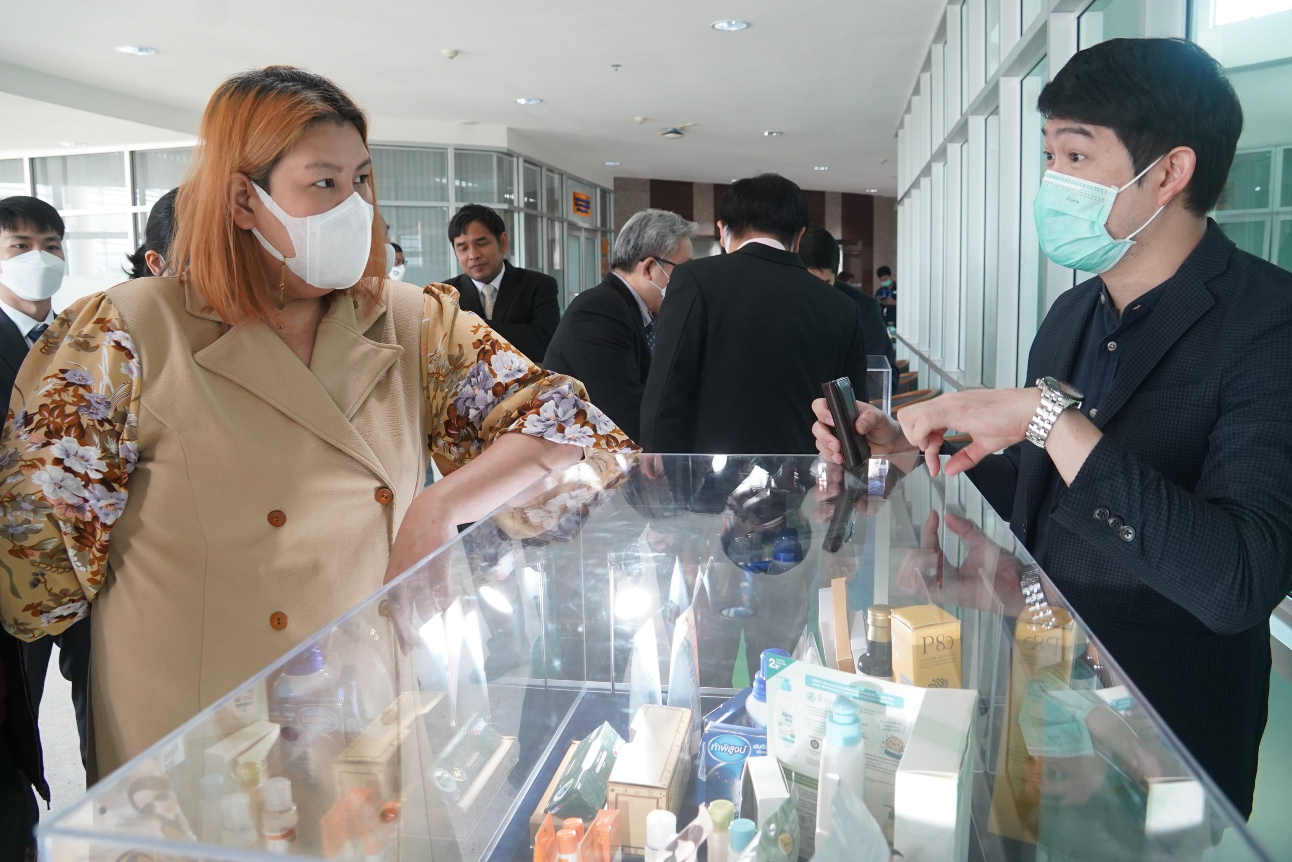
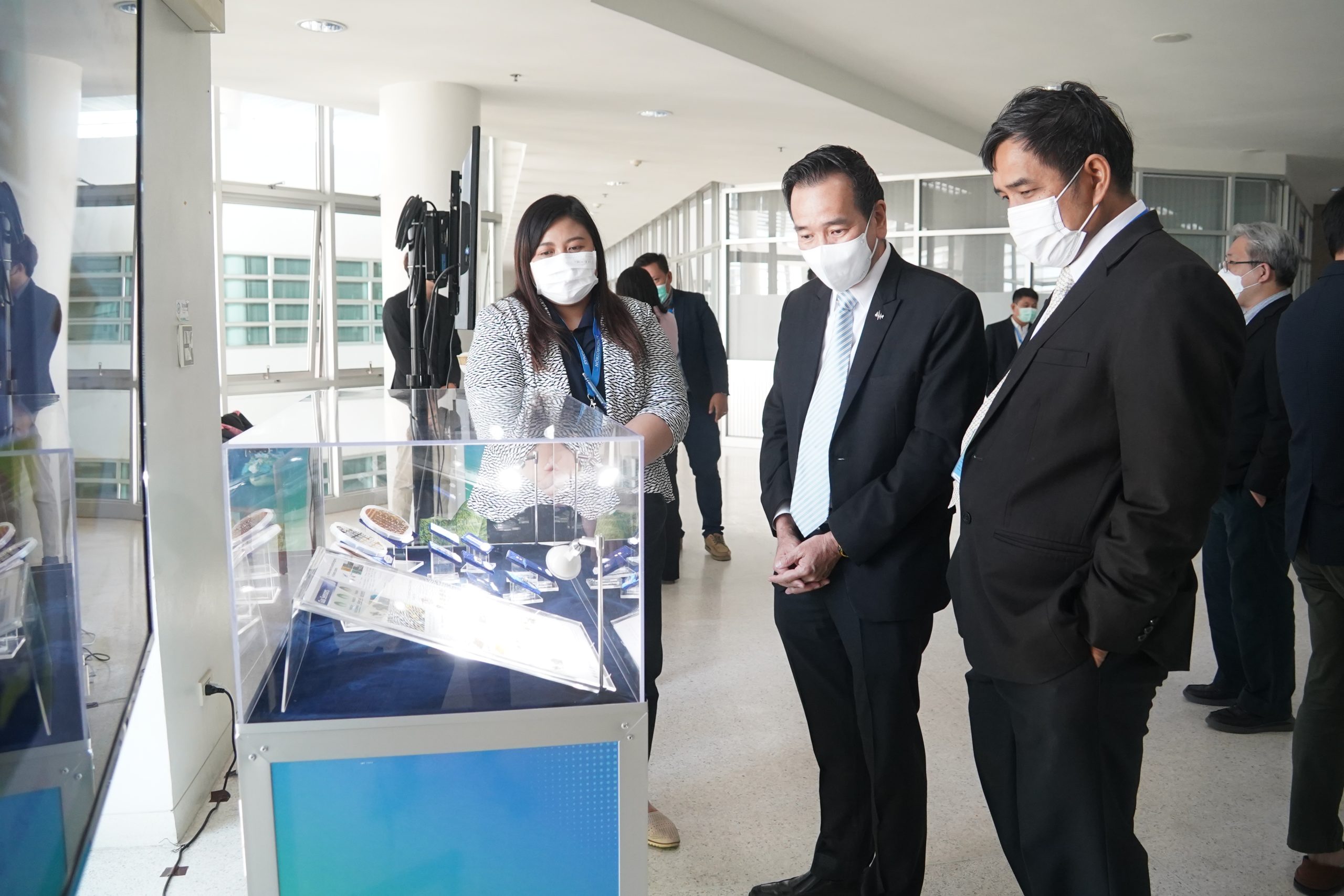
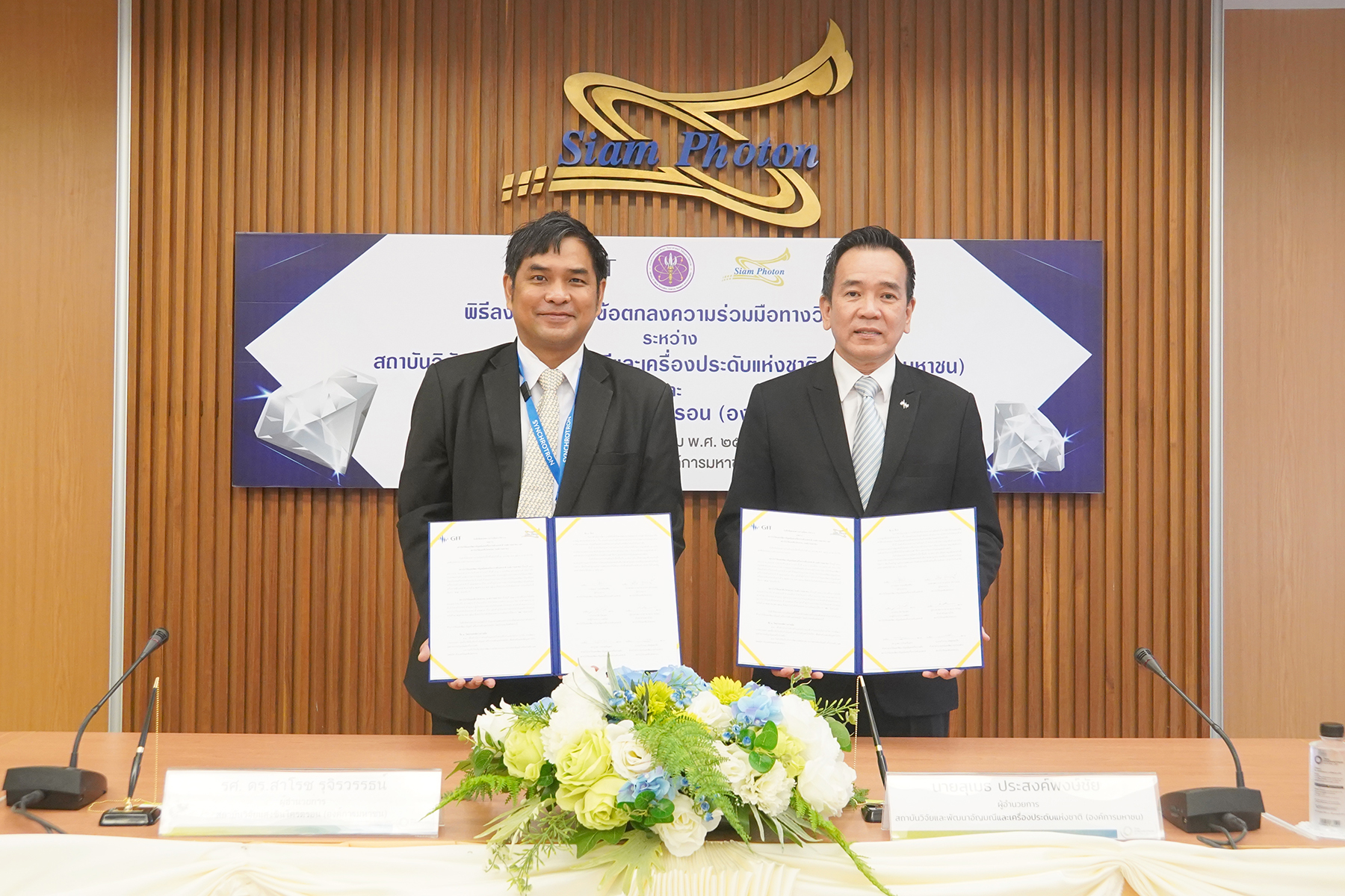
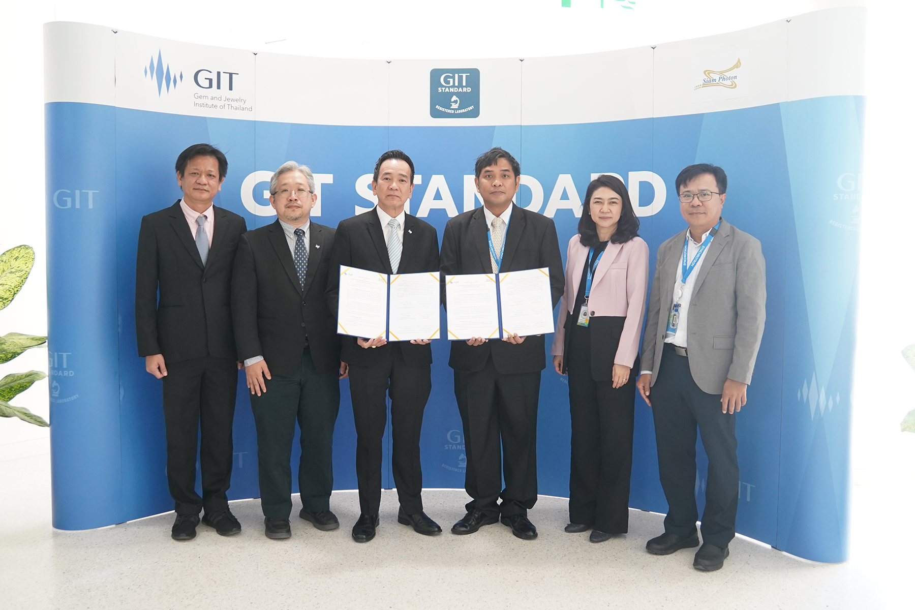
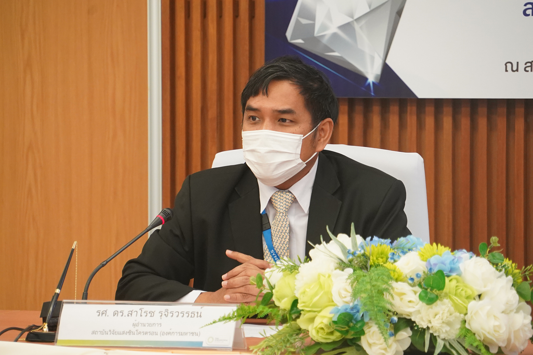
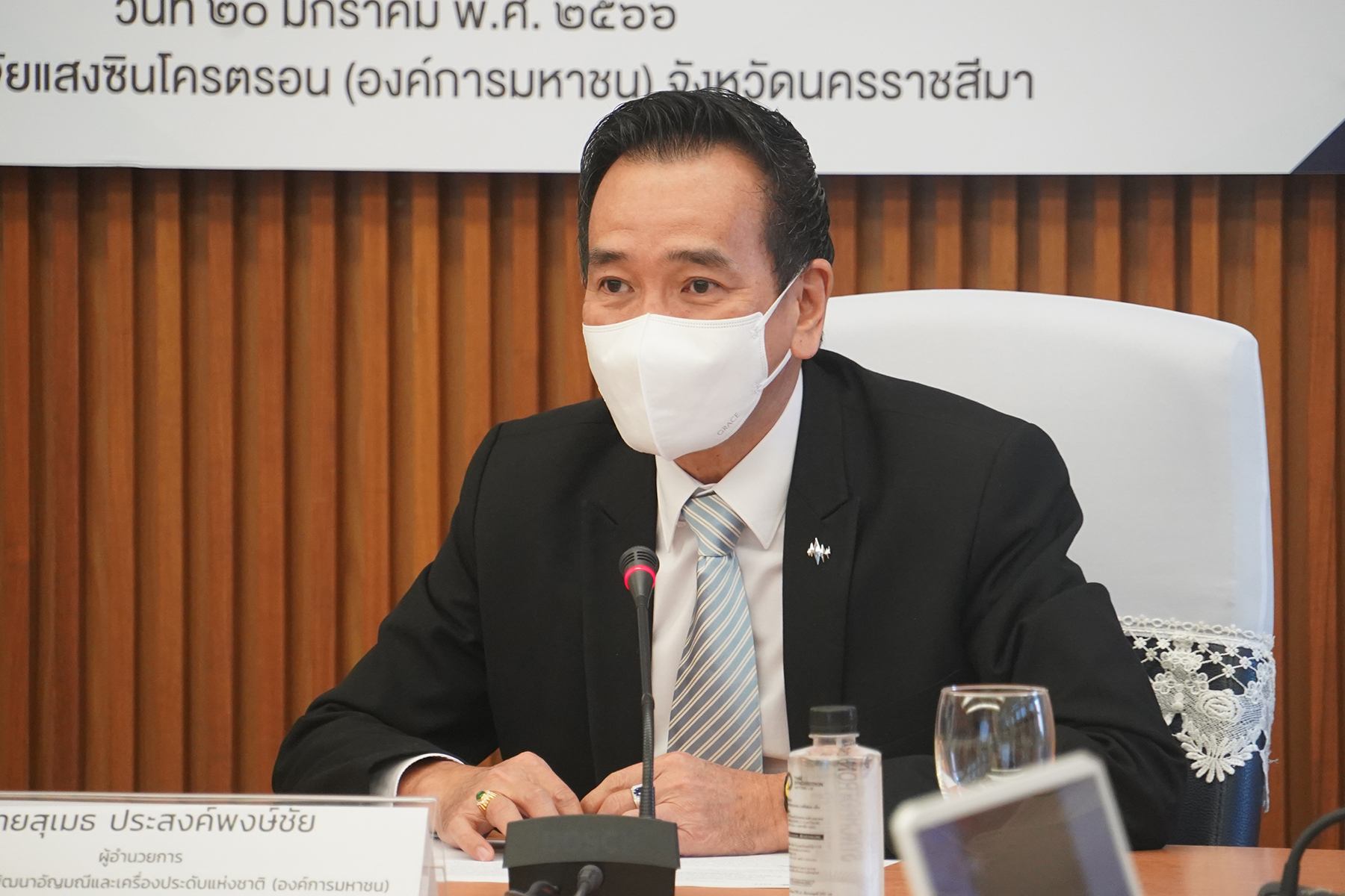
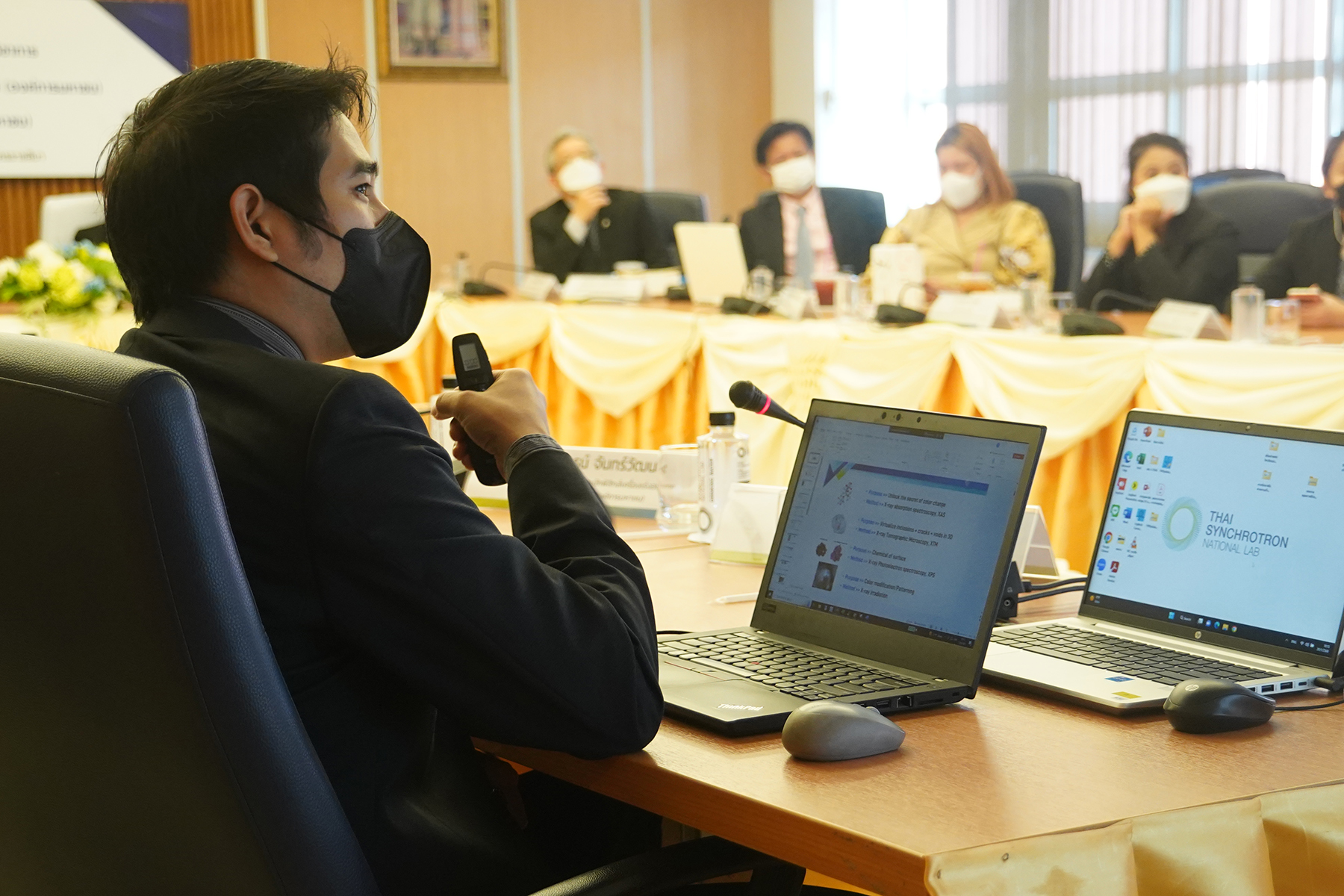
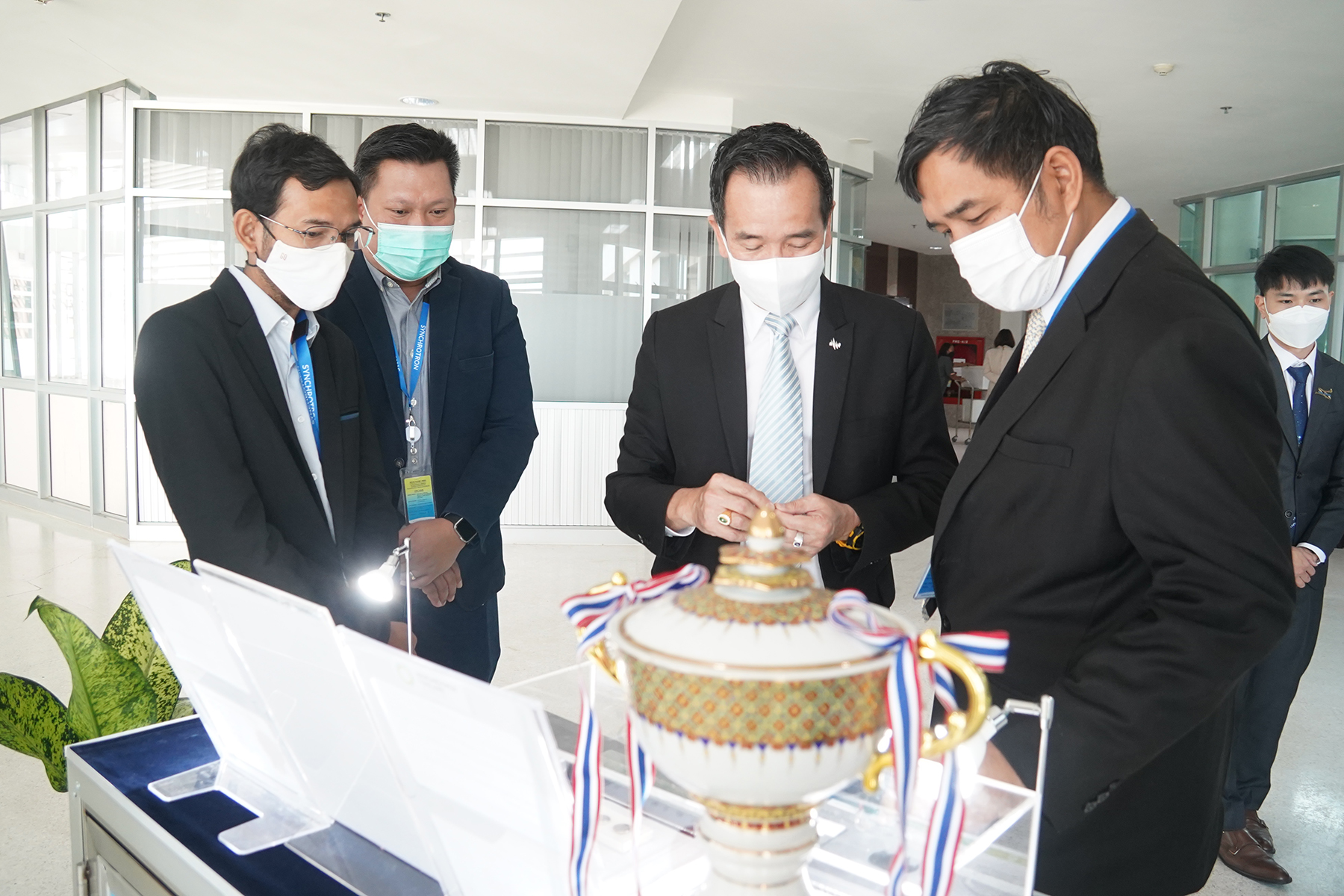
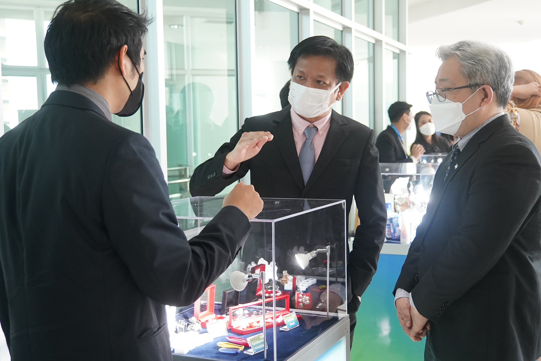
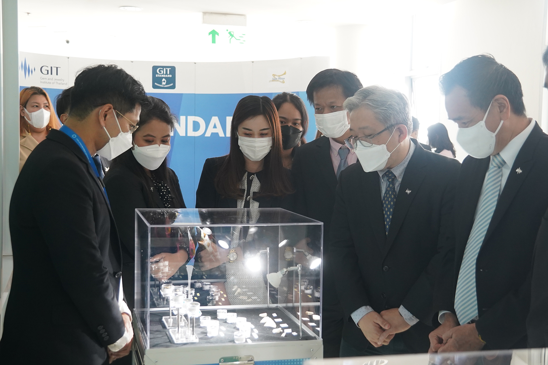
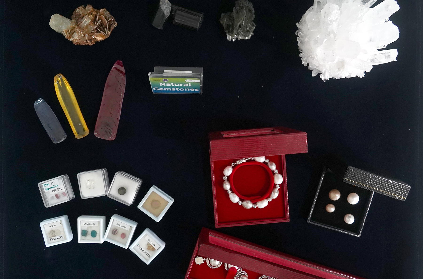
On 20 January 2023, Assoc. Prof. Dr. Saroj Rujirawat, Director of Synchrotron Light Research Institute (Public Organization) or SLRI, Ministry of Higher Education, Science, Research and Innovation, and Mr. Sumed Prasongpongchai, Director of The Gem and Jewelry Institute of Thailand (Public Organization) or GIT signed the Memorandum of Understanding (MoU) at SLRI in Nakhon Ratchasima to promote SLRI-GIT collaboration on research and development of gems, jewelry, and precious metals by applying synchrotron light and related technologies. The collaboration is expected to increase value of gem and jewelry industry and make Thailand the world’s gem and jewelry trading and manufacturing hub.
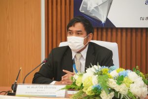
Assoc. Prof. Dr. Rujirawat said, “SLRI and GIT intend to build academic collaboration between the two institutes to foster research implementation, quality improvement and development, testing, and other activities related to gems, jewelry, and precious metals by applying synchrotron light and related technologies. The collaboration will increase value of gem and jewelry industry. The two institutes also aim to develop our personnel, promote knowledge exchange, and build strong research network between government organizations. Other activities deemed appropriate by both SLRI and GIT will be organized as well.”
Mr. Prasongpongchai stated, “SLRI and GIT will not only jointly promote research and development of testing process technique and quality improvement processes of gems, jewelry, and precious metals but also provide each other with equipment for research, analysis, testing, and inspection. The two institutes will also support each other in applying synchrotron light and related technologies in using equipment, tools, laboratories, and other factors related to research implementation. Moreover, we will foster trainings and exchange of our personnel as well as perform knowledge transfer related to research to develop skills, performance, and capacity of our scientists which account for country development.”
Mr. Prasongpongchai added, “I do hope that this MoU will lead to our collaboration for making Thailand the world’s hub for gem and jewelry trade and manufacture in all aspects.”
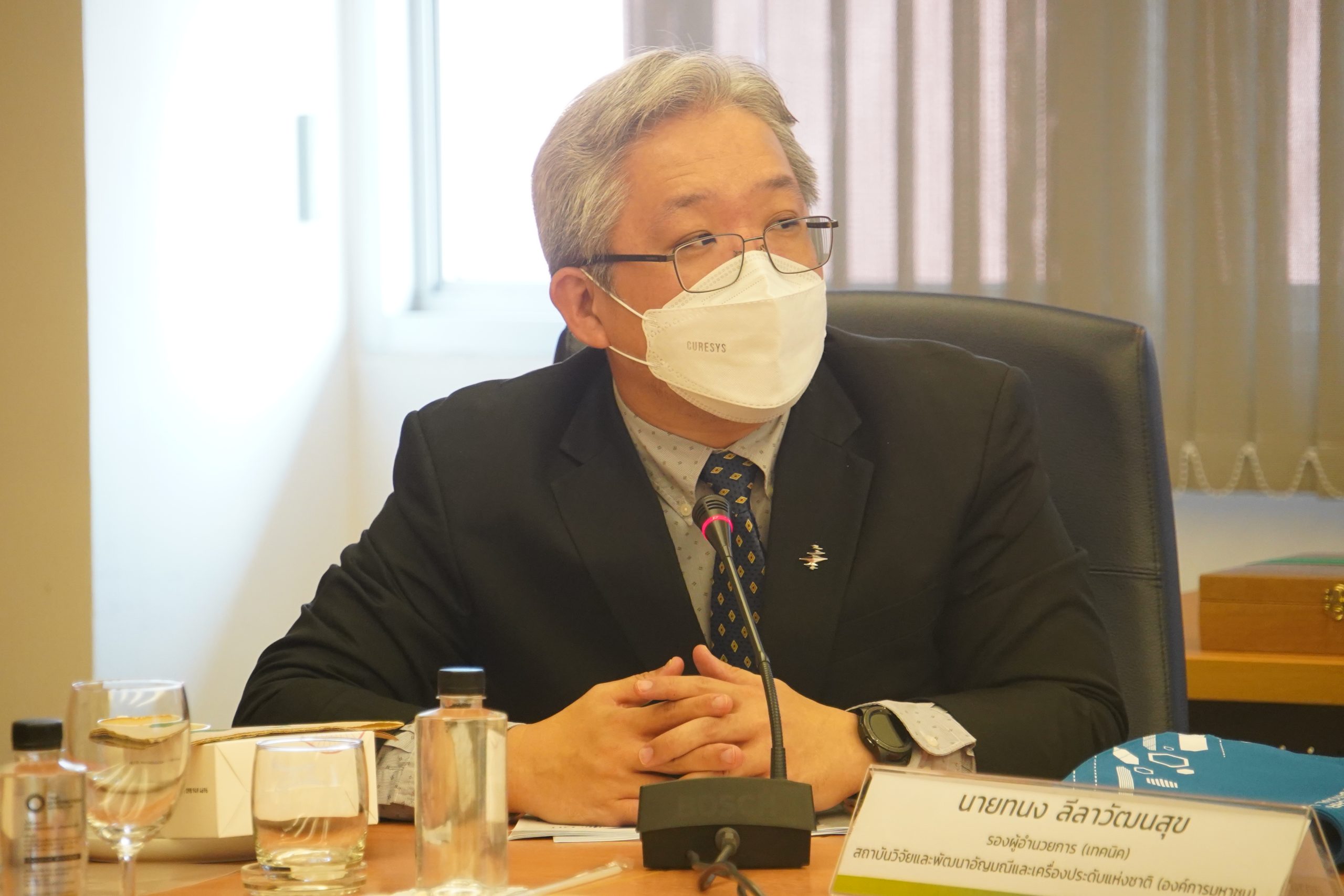
On this occasion, special lectures were provided by both GIT and SLRI experts for knowledge exchange. Dr. Chatree Saiyasombat, Acting Beamline Manager, BL.7.2W, SLRI gave lecture under the topic “Synchrotron light application and research on gems” while Mr. Thanong Leelawatanasuk, deputy director on technique and Dr. Bhuwadol Wanthanachaisaeng, chief of gem and jewelry research and development section, GIT provided lecture on “Guidelines for gem and jewelry research in Thailand.”
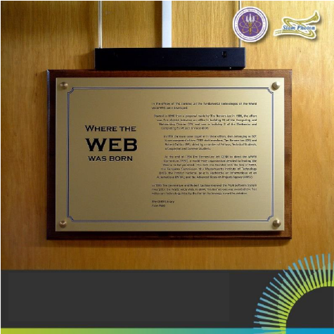
Do you know that the World Wide Web(WWW) information system used in daily life was invented by the world’s most famous research institute called European Organization for Nuclear Research or CERN? This could make many people wonder why WWW was created by a physics laboratory rather than a computer organization or renowned company.
CERN is the largest particle physics laboratory on earth located at border between French Republic and Swiss Confederation. The laboratory possesses scientific equipment for studying particle physics called a particle accelerator and has built the most advanced particle accelerator network ever. The accelerators increase energy and speed of proton or lead particles to nearly speed of light before collision with each other. New types of particles that are created after the collision as predicted by the particle physics theory will be studied by researchers.
Due to the large number and varieties of particles created, it is very challenging, difficult, and important for researchers to store them properly before starting to extract these data for investigation.
For effectiveness of data sharing among researchers, Mr. Tim Bernes-Lee launched in 1980 the project called ENQUIRE to develop a program which runs by using hypertext messages. In 1990, he collaborated with Mr. Robert Cailliau in developing a data management system with higher quality under the project called “WorldWideWeb” (“W3” or “Web” in short). The system is a data network linked by using hypertext and displayed on the program called browser. Many browsers are used at present such as Google Chrome, Safari, Firefox, etc.
The Web was launched for official use in 1991 and in 1993 CERN announced that everyone has free access to the Web. The data network has been continually developed ever since and use of the Web by the public began in 1993-1994.
The Web development may not be considered among CERN’s main missions, but obstacles and challenges CERN researchers face in storing and sharing huge experiment data have led to the creation of related technology for application in our daily lives.
Synchrotron Light Research Institute (Public Organization) has so far collaborated with CERN in the development of an internal particle tracking system containing pixel sensors as one of the most important components. The sensors generate electric signal when hit by high-energy particles. Not only is this type of sensor used in physics experiment, but also in taking X-ray tomography for medical purpose.
Article by:
Dr. Kritsada Kittimanapun, Accelerator Physicist, SLRI