About Beamline
BL5.2: SUT-NANOTEC-SLRI XAS Beamline
BL5.2 has been constructed for the project ‘SUT-NANOTEC Joint Research Facilities for Synchrotron Utilization’ at the Synchrotron Light Research Institute. The scientists and researchers from these three organizations (SUT, Nanotec and SLRI) will work together in scientific research and development with synchrotron radiation.
SUT-NANOTEC-SLRI beamline (BL5.2) is dedicated to X-ray Absorption Spectroscopy (XAS)technique. It can be used to determine chemical speciation and local structure (type of neighboring atoms, coordination number, inter-atomic distance) of the absorbing atom. Moreover XAS is a non-destructive tool which can be carried out on any type of material, e.g. solids (crystalline or amorphous), liquids and gases, under non-ambiant conditions (pressure and temperature control).Consequently, can be employed to study samples in different scientific areas, such as materials science, biology, environmental science, archeology and geology.
An Example of Sulfur Speciation by Sulfur K-edge XANES Spectroscopy
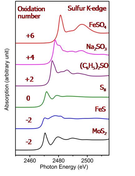
Fig.1 The S K-edge XANES data acquired at BL5.2
X-ray Absorption Near Edge Structure (XANES) spectroscopy is an ideal non-destructive technique for characterizing and quantifying S species in compositionally complex materials such as minerals, coals, soils and rubber. Shifts in the position of the absorption-edge feature of S K-edge XANES spectra can be applied for determining the oxidation state of both inorganic and organic species of sulfur. XANES spectra can be also used as fingerprints to identify the coordination chemistry.
BL5.2 uses a design similar to BL8 with an in-house fabricated fixed-exit double crystal monochromator (DCM). The x-ray energy is tunable by a DCM equipped with several types of crystal for covering photon energy from 1240 eV to 12100 eV. K-edge absorption of Magnesium up to Gallium can be studied. Other heavier atomic species can be investigated via L or M edges. BL5.2 was designed by engineer and scientist team at SLRI. Most of components were in-house fabricated. The beamline has been installed and commissioned. It is available for users since March 2013.
Since BL5.2 is a joint project between SUT, Nanotec and SLRI, BL5.2 beamtime will be provided equally to these three organizations. A major part of SLRI beamtime will be given to general users who submit their beamtime applications via our website.
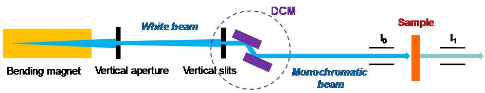
Fig.2 Schematic of SUT-NANOTEC-SLRI beamline (BL5.2)
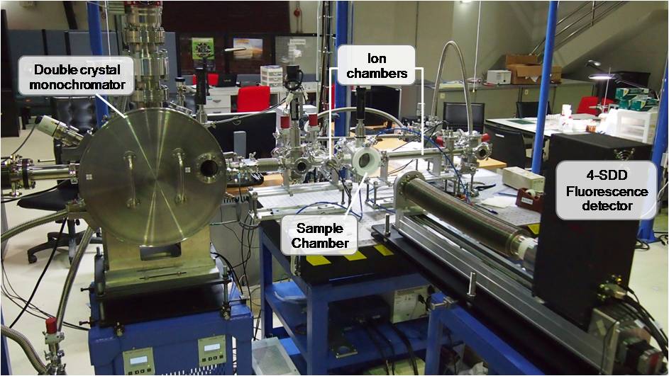
Fig.3 Current SUT-NANOTEC-SLRI beamline setup (BL5.2)
Beamline Characteristics
| Energy range | 1810 eV-13000 eV |
| Beam size at the sample |
- 13 mm (width) × 1 mm (height) for Transmission mode - 20 mm (width) × 1 mm (height) for Fluorescence mode |
| Flux | 108-1010 photons/sec/100 mA |
| Energy resolution | 2 × 10⁻⁴ |
| Experimental Setup | - Transmission mode with Ion chambers
- Fluorescence mode with 4 element Silicon drift detector |
| Crystal type | InSb(111) : 2d spacing (Å) = 7.481
: Energy rang = 1830 - 7000 eV Ge(220) : 2d spacing (Å) = 4.001 : Energy rang = 3440 - 12100 eV |
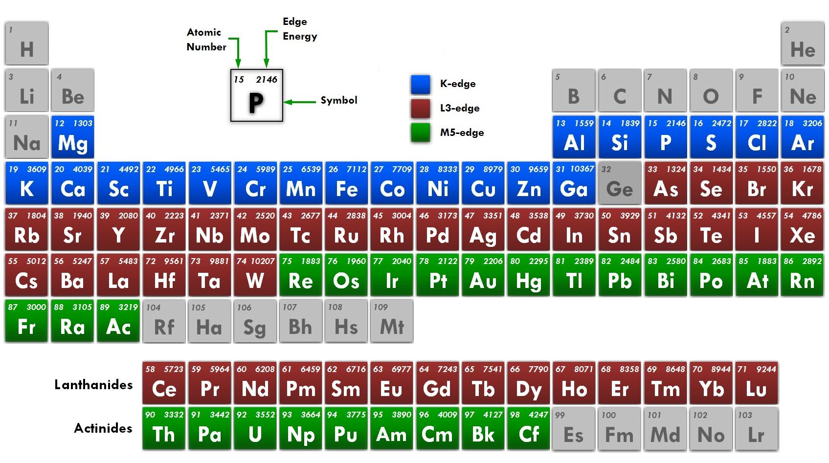
Fig. 4 Shows the absortion edges that can be measured at BL5.2
* In the case of Ga , the element can be measured spectrum depends on the Concentration of Ga atoms in the sample.
Commissioning results of SUT-NANOTEC-SLRI beamline setup (BL5.2)The beamline has been installed and checked the quality of XAS spectra at low energy (S K-edge, about 2472 eV) and high energy (Cu K-edge, about 8979 eV)
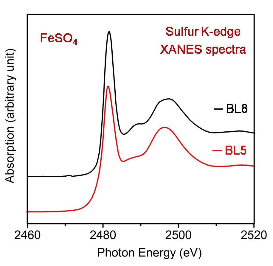
Fig.5 Comparison of S K-edge XANES spectra ofFe2SO4measured atBL5.2 andBL8
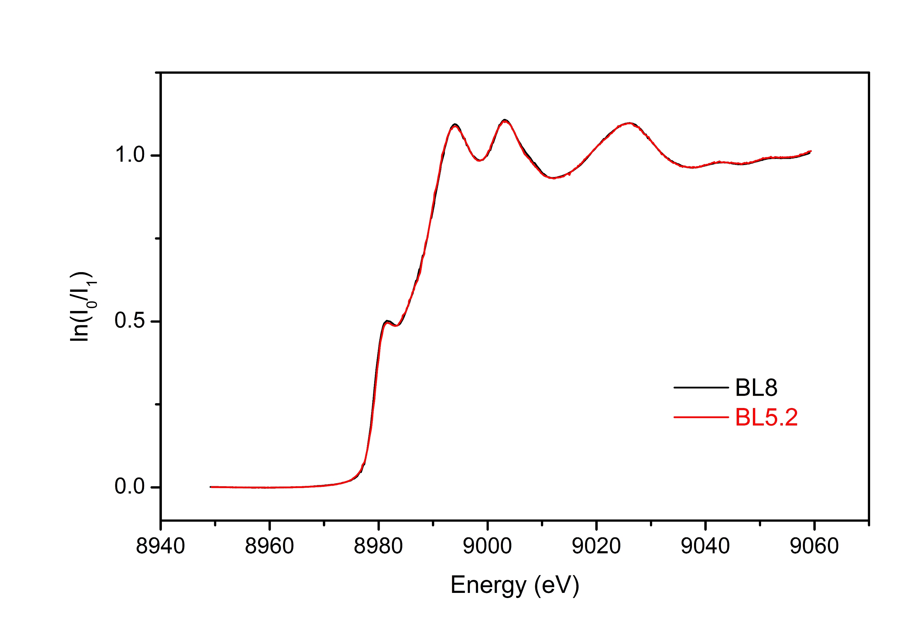
Fig.6 Comparison of Cu K-edge XANES spectra ofCu foilmeasured atBL5.2 andBL8
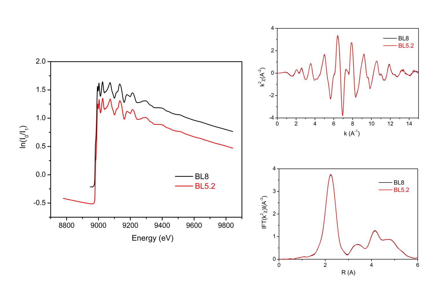
Fig.7 Comparison of Cu K-edge EXAFS spectra ofCu foilmeasured atBL5.2 andBL8
Moreover, XAS spectra of diluted sample have been measured in fluorescence mode by using 4-element silicon drift detector at BL5.2
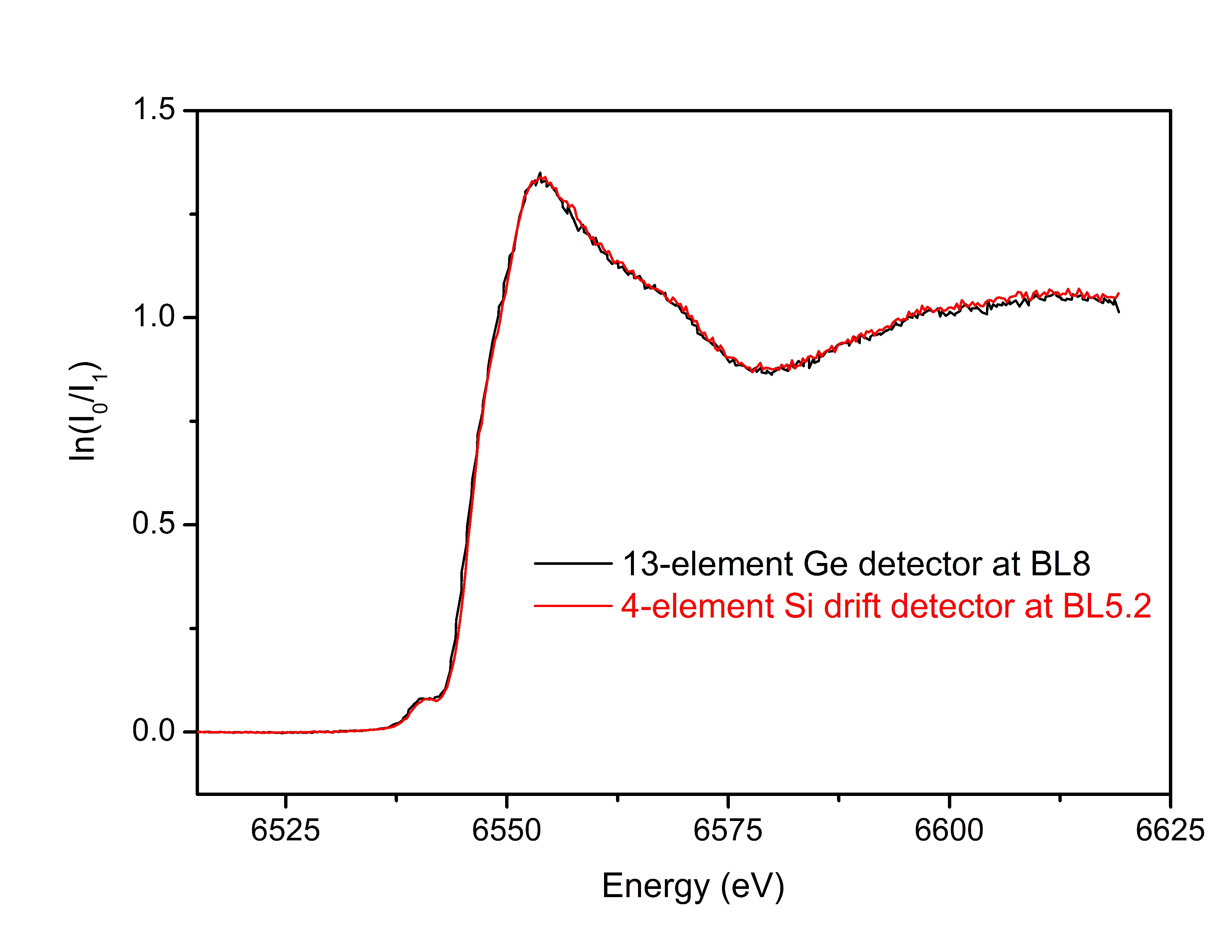
Fig.8 Comparison of Mn K-edge XANES spectra of a diluted sample (Mn content of
1.6 %wt) at BL5.2 and BL8


111 SirinthonWitchothai Building, University Avenue, Muang District, Nakhon Ratchasima 30000, THAILAND
Tel.: +66 44 217 040 Fax: +66 44 217 047 Email: siampl@slri.or.th
Copyright © 2015 Synchrotron light research institute. All Rights Reserved.
|
|