Beamline
A beamline is a system of components, such as slits, monochromators, focusing mirrors and other optical elements used for shaping and delivering the synchrotron light from the storage ring to the experimental stations. An experimental station, located at the end of a beamline, contains measurement systems for a specific measurement technique. SPL currently has 10 beamlines with 12 experimental stations in routine operation and 3 beamlines and 4 experimental stations under construction.
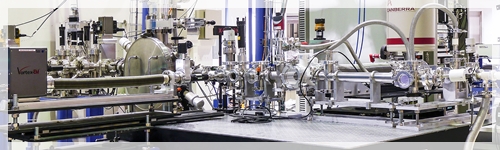
1. BL1.1W: Multiple X-ray Techniques (MXT)
The experimental techniques available are x-ray absorption spectroscopy (XAS) and x-ray fluorescence (XRF). The XAS can be used to determine oxidation states and atomic scale structure of materials. The XRF technique is used to qualitatively and quantitatively determine elemental compositions of materials. Experiments employing any of the techniques or coupling of the two techniques could be included in the proposal.
X-ray diffraction (XRD) technique will be added to the beamline capability in 2018. The technique is used to study crystal structures.
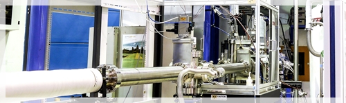
2. BL1.2W: X-ray Imaging and X-ray Tomographic Microscopy (XTM).
Beamline 1.2w is an X-ray beamline dedicated to X-ray imaging and computed microtomography. The 3D reconstructed images produced from BL1.2W will allow for non-destructive visualization and investigation of the matter inside a sample.
3. BL1.3 W: Small/Wide Angle X-ray Scattering (SAXS/WAXS)
Beamline 1.3W is dedicated for nano structural investigation of material such as analysis of particle size and size distribution, period of block copolymer, starch lamellar nano structure, percent crystallinity etc
4. BL2.2: Time-resolved X-ray Absorption Spectroscopy (TRXAS)
The short data collection time of TRXAS beamline opens a new opportunity for in situ x-ray absorption spectroscopy (XAS) experiments such as studies of changes of the electronic structures or the local coordination environments of an atom during a change in thermodynamic conditions.
5. BL3.2Ua/b: PES/PEEM
BL3.2Ua: PES (Photoelectron Emission Spectroscopy)

Soft-X ray spectroscopy beamline analyzes atomic elements and its chemical states of surface and interface of materials.
BL3.2Ub: PEEM (Photoelectron Emission Microscopy)
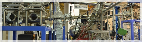
The PEEM end station produces high resolution images from electrons that are emitted from the samples by the excitation with synchrotron soft X-ray. Additionally, information related to the surface chemistries and electronic structures of sub-micron domains in the images can be obtained via spatially-resolved near-edge X-ray absorption spectroscopy.
6. BL4.1: Infrared Spectroscopy and Imaging (ISI)
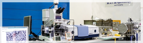
Fourier Transform Infrared (FTIR) spectroscopy has been widely used for a routine analytical technique. Thanks to the high brightness of synchrotron radiation, synchrotron based Infrared microspectroscopy can provide high spatial resolution, good signal to noise ratio and short data acquisition time. (Beamline is under construction, experimental station is in operation with conventional IR source)
7. BL5.2: X-ray Absorption Spectroscopy (XAS), SUT-NANOTEC-SLRI XAS Beamline
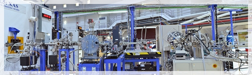
Beamline 5.2 is one of Synchrotron Radiation beamline at SLRI which dedicated to an X-ray Absorption Spectroscopy (XAS). This beamline is used to study an atomic arrangement and elemental speciations of materials. BL5 is a consortium beamline, jointly funded by Suranaree University of Technology, the National Nanotechnology Center and SLRI
8. BL6a/b: DXL/Micro-XRF
BL6a: DXL (Deep X-ray Lithography)
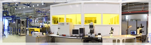
Beamline 6a is a micromachining beamline using Deep X-ray Lithography (DXL) for fabrication of micro mechanical components.
BL6b: Micro-XRF (Micro-X-ray Fluorescence)
Beamline BL6b Micro X-ray Fluorescence (µ-XRF) Spectroscopy/Imaging
- To detect trace elements in materials such as plants, soil.
- To determine the elemental compositions in materials such as metal oxide compounds
- To study the elemental distributions in inhomogeneous materials
9. BL7.2W: Macromolecular Crystallography (MX)
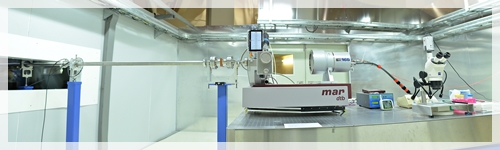
Beamline 7.2W is a tunable energy beamline dedicated to macromolecular crystallography for determining the crystal structure of proteins, their complexes and other molecules at atomic resolution.
10. BL8: X-ray Absorption Spectroscopy (XAS)
Beamline 8 is a X-ray Absorption Spectroscopy (XAS) beamline for studies of atomic arrangement in material


111 SirinthonWitchothai Building, University Avenue, Muang District, Nakhon Ratchasima 30000, THAILAND
Tel.: +66 44 217 040 Fax: +66 44 217 047 Email: siampl@slri.or.th
Copyright © 2015 Synchrotron light research institute. All Rights Reserved.
|
|