BL3.2b: PEEM
At BL3.2b (PEEM), a sample is irradiated with monochromatic light from varied line-spacing plane-grating monochromator. Electrons which are created by photoemission and photoabsorption processes are projected by a set of magnetic lenses onto a micro-channelplate intensifier and a phosphor screen where the final image forms. By scanning incident photon energy and capturing PEEM image at each energy step, a series of images which contains photoabsorption spectra from nanometer-sized areas of the sample can be obtained. Alternatively, photon energy is fixed and an electron energy analyzer is used to determine the photoemission spectra of photoelectrons emitted from certain areas of the sample.
|
Technical information |
|
|
Technique: |
Photoemission Electron Microscopy (PEEM) |
|
Applications: |
Electron microscopy for surface, interface and thin-film researches. |
|
Source and beamline |
|
|
Radiation source : |
U-60 undulator |
|
Photon flux at sample: |
1e12 phs/s at 100 eV |
|
Photon energy range: |
40-160eV and 220-1040 eV |
|
Monochromator type: |
Varied-line-spacing plane-grating monochromator |
|
Energy bandwidth: |
0.01% minimum |
|
Experimental Station |
|
|
Model: |
Elmitec PEEM III |
|
Image resolution: |
30 nm |
|
Sources: |
Synchrotron, Hg UV lamp, electron gun for low-energy electron microscopy (LEEM) |
|
Samples: |
Must be flat and smooth, ultra-high vacuum compatible, and electrically conductive. Sample holder for flat and thin samples can heat up to > 1500°C. Non-conductive samples can be coated with metal films before imaging. |
|
Detectors: |
Micro-channel plate, Sensicam QE CCD camera |
|
Analyzer: |
Hemispherical electron energy analyzer |
|
Sample environment: |
Ultra-high vacuum. Base pressure at 10e-10 torr. |
|
Beamline status: |
Commissioning |
Beamline layout
.jpg)
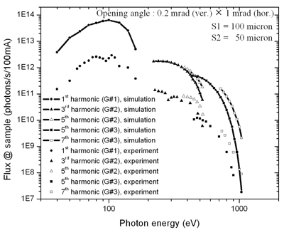
Photon flux from BL-3 at PEEM’s sample position
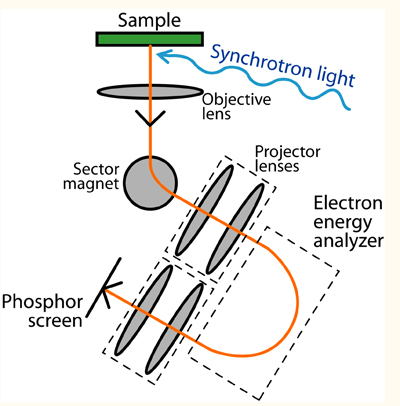
Schematics of PEEM system
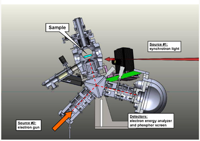
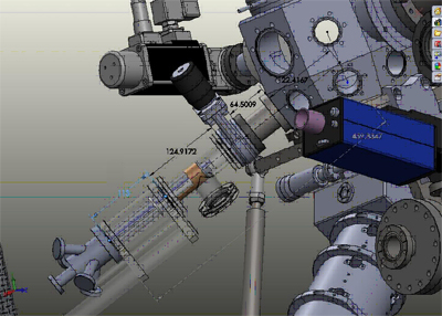
Mechanical design of PEEM/LEEM system
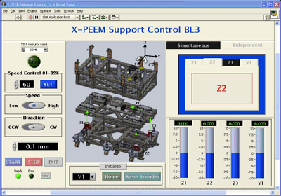
Computer software that controls PEEM mechanical support for alignment with synchrotron beam
Thin-film deposition system can be installed at PEEM’s experimental chamber for in-situ observation of thin-film growths
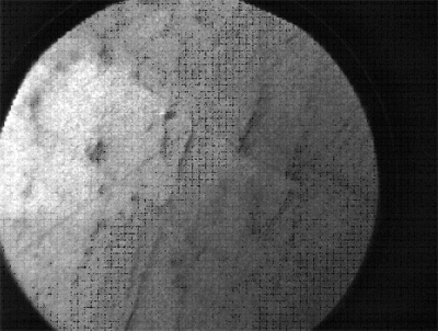
PEEM image of tungsten. Field of view is 75 microns.
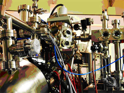
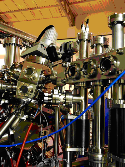
PEEM system
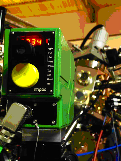
PEEM sample can be heated up to > 1500°C. The temperature can be measured using an infrared pyrometer. PEEM can observe the sample during temperature change in real-time.
The loadlock of PEEM can store more than 4 samples in vacuum. Samples can be annealed or deposited with thin films by various methods.


111 SirinthonWitchothai Building, University Avenue, Muang District, Nakhon Ratchasima 30000, THAILAND
Tel.: +66 44 217 040 Fax: +66 44 217 047 Email: siampl@slri.or.th
Copyright © 2015 Synchrotron light research institute. All Rights Reserved.
|
|