



ภาพรวมระบบลำเลียงแสงและสถานีทดลอง
A beamline is a system of components, such as slits, monochromators, focusing mirrors, and other optical elements used for shaping and delivering the synchrotron light from the storage ring to the experimental stations. An experimental station, located at the end of a beamline, contains measurement systems for a specific measurement technique. The SLRI beamline currently has 11 beamlines with 13 experimental stations in routine operation.
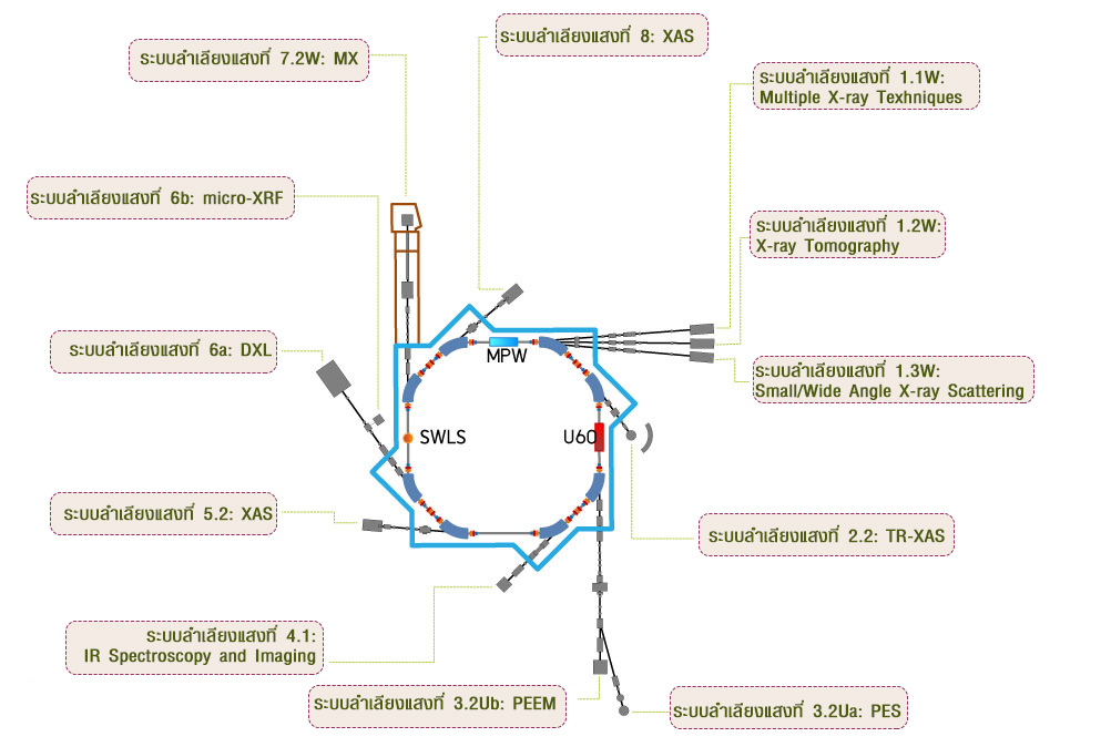
Beamline Map
ระบบลำเลียงแสงที่แยกออกจากวงกักเก็บอิเล็กตรอน SPSกดที่ชื่อสถานี (BL) บนแผนที่ เพื่อดูเทคนิคที่ให้บริการและลิงก์เว็บไซต์ของสถานี
Beamline List
รายชื่อสถานีทดลอง 13 สถานี · กดเพื่อดูรายละเอียดAdvanced Techniques at SLRI
Powerful methods for science and technology · กดชื่อ BL เพื่อดูสถานีที่ให้บริการSpectroscopy19 เทคนิค
Investigates local atomic structure and electronic states by analyzing X-ray absorption patterns.
Captures dynamic changes in electronic and structural properties over time.
Combines XAS with micro-focused beams for localized structural analysis.
Surface-sensitive absorption spectroscopy for thin films and interfaces.
Determines elemental composition by detecting characteristic X-ray emissions.
Ultra-trace elemental analysis using total reflection geometry with very low detection limits.
Identifies chemical bonds and functional groups using infrared absorption spectra.
Analyzes electronic structure by detecting photoelectrons.
Quantifies elemental composition and chemical states within the top nanometers via core-level photoemission.
Depth-sensitive XPS using emission angle to separate surface and near-surface information and probe anisotropy.
Studies electronic structure by measuring photoelectrons emitted from a sample.
Probes electronic band structure by measuring photoelectrons emitted at different angles.
Studying orientation-dependent absorption.
Near-edge region of XAS sensitive to bonding, functional groups, and molecular orientation.
Spatially resolved XPS providing micron-scale chemical state mapping for surfaces.
Probes the electronic band structure with high spatial resolution, enabling microscale analysis.
Electron Energy Loss Spectroscopy at microscopic scale to probe electronic structure and chemical composition.
High-resolution NEXAFS imaging for mapping molecular orientation and bonding.
NEXAFS detected via Auger yield with micron spatial resolution for chemical state mapping.
Diffraction7 เทคนิค
Identifies crystalline phases in powdered samples by analyzing diffraction patterns.
A cutting-edge method for probing site-specific atomic arrangements and subtle structural variations in complex materials.
Determines 3D structures of macromolecules by analyzing X-ray diffraction patterns from crystals.
Determines residual stress in materials by measuring diffraction at different tilt angles.
Surface diffraction technique using low-energy electrons to determine atomic structure and surface symmetry.
Localized LEED analysis with micrometer-scale resolution for studying specific surface regions and defects.
Quantitative surface diffraction technique analyzing LEED intensity as a function of electron energy to determine atomic structure of surfaces.
Scattering4 เทคนิค
Characterizes nanoscale structures and particle size distributions.
Characterizes crystalline order and structural motifs at shorter length scales.
Surface-sensitive scattering technique for thin films and nanostructures.
Determines thickness, density, and roughness of thin films by analyzing reflected X-rays.
Microscopy3 เทคนิค
Full-field electron microscopy technique using photoemission to visualize surface morphology, electronic structure, and chemical contrast at the nanoscale.
Real-time, surface-sensitive imaging using elastically backscattered low-energy electrons to visualize phases, domains, and reconstructions.
Acquires intensity–voltage (I–V) curves of diffracted electron beams to determine surface atomic structure with high precision.
Imaging7 เทคนิค
Non-destructive technique for elemental analysis and mapping using focused X-rays at micrometer-scale resolution.
Maps elemental distribution in a sample using X-ray fluorescence over a wide field.
3D imaging technique using X-rays to reconstruct internal structures with micrometer resolution.
Uses X-rays to create two-dimensional images of internal structures with high resolution.
Visualizes chemical bonding and molecular orientation at surfaces using NEXAFS imaging.
Chemical-state mapping over fields of view using scanning or full-field XPS detection.
Full-field photoemission imaging to spatially resolve work-function and electronic contrast at the nanoscale.
Lithography1 เทคนิค
A microfabrication method using high-energy X-rays to produce high-aspect-ratio structures for MEMS and microfluidics.
Support Facilities & Equipment
Siam Photon Laboratory (SPL) has prepared a range of advanced instruments and facilities to support usersBasic tools for sample preparation
- Pestle and mortar
- Tweezers
- Spatulas, etc.
Readability: 0.01 g
- Built-in glass draft shield for high weighing stability.
- Applications: general laboratory use, formulation, and quality control where 0.01 g precision is required.
Readability: 0.1 mg (0.0001 g)
- Built-in glass draft shield for high weighing stability.
- Applications: high-precision weighing for analytical chemistry, sample preparation, and pharmaceutical research.
Chemical fume hood
- Work area: 1200 mm (W) × 800 mm (D) × 1200 mm (H)
- Airflow velocity: 0.5 m/s (average face velocity at sash opening)
- LED lighting, ≥ 800 lux inside work area
Pressing pellets
- Maximum force: 15 US tons (13.6 metric tons)
- Evacuable pellet die diameters: 6, 10, 13, 16 and 20 mm
- Each die set has a maximum force rating that should not be exceeded — check the front of the die for the maximum load capacity.
Controlled heating for lab processes
- Volume: 53 liters (1.9 cu. ft.)
- Working temp. range: ambient +10 °C to +300 °C
- Temperature accuracy: 0.1 °C up to 99.9 °C, 0.5 °C from 100 °C
- Adjustable temperature, fan speed, air flap position and program time
Extremely fast grinding of very small samples in a single step
- Adjustable from 900 to 3,000 oscillations/min (15–50 Hz)
- Max feed size 6 mm · final fineness down to 5 µm
- Sample quantity 0.1 – 5 ml
- Bowls 5, 10, 15 ml · balls 0.1 – 15 mm · zirconium oxide, stainless steel, PTFE
How to Apply for Beamtime
ขั้นตอนการขอใช้เวลาแสงซินโครตรอนSign in to the Beamtime Application System (BeamApp) to submit your proposal.
Sign InDownload the beamtime application procedure manual.
Download Manual (PDF)All users must strictly follow the safety protocols for beamline operation.
อ่านแนวปฏิบัติEstimate Your Beamline Shifts
To view detailed information about shift estimation, please return to the homepage of the beamline you are interested in. Each beamline provides its own specific guidelines and data for calculating shifts.
Safety Guidelines for Beamline Operation
To ensure safe and compliant beamline operations, all users must strictly follow the established safety protocols.- Only authorized personnel are permitted to operate or control the beamline equipment.
- All users must complete the Beamline Hutch Search and Secure training, as well as radiation safety procedures, before operating any beamline system.
- Users are also required to sign the form in Appendix 4: Beamline Safety Training.
- Users are not allowed to remain in the experimental hall during beam injection under any circumstances.
- A radiation monitoring badge must be worn at all times while inside the experimental hall.
- Food and beverages are strictly prohibited at the beamline area.
- Users are responsible for cleaning and tidying the beamline area on the final day of their scheduled beamtime.
- Users must take back their samples, chemicals, and any chemical waste generated during experiments.
- End of Experiment Feedback: complete the post-experiment satisfaction evaluation through the BeamApp system once your experiment has concluded.
- Experimental Report: submit a summary report of your experimental results within 14 days after the completion of your experiment via BeamApp.
Publications
Explore published research outputs by fiscal year through an interactive Power BI dashboard.Multiple X-ray Techniques
MXT · Established 2016
Service Techniques
XAS / GIXAS / PXRD / AXRD / WAXS / GIWAXS / XRR / XRF / Residue Stress (Sin²Ψ method)
รายละเอียดเทคนิค (กดเพื่อดู)
X-ray Imaging & Microtomography
XTM · Established 2016
Service Techniques
XTM / X-Ray 2D Imaging
รายละเอียดเทคนิค (กดเพื่อดู)
Small & Wide-Angle X-ray Scattering
SAXS/WAXS · Established 2011
Service Techniques
SAXS / WAXS / XRF
รายละเอียดเทคนิค (กดเพื่อดู)
Time-resolved X-ray Absorption Spectroscopy
TRXAS · Established 2011
Service Techniques
TRXAS
รายละเอียดเทคนิค (กดเพื่อดู)
X-ray Photoelectron Spectroscopy (SUT-NANOTEC-SLRI)
XPS · Established 2011
Service Techniques
XPS for SUT-NANOTEC-SLRI
รายละเอียดเทคนิค (กดเพื่อดู)
Photoelectron Emission Spectroscopy
PES · Established 2003
Service Techniques
PES / ARPES / UPS / XPS / ARXPS / NEXAFS / ARXAS
รายละเอียดเทคนิค (กดเพื่อดู)
Photoemission Electron Microscopy
PEEM · Established 2011
Service Techniques
PEEM / LEEM / LEEM-IV / micro-LEED / LEED-IV / micro-EELS / ARPES / micro-ARPES / micro-XPS / micro-NEXAFS / Auger yield micro-NEXAFS / LEED / Imaging-XPS / Imaging NEXAFS / Work function mapping
รายละเอียดเทคนิค (กดเพื่อดู)
micro-Infrared Spectroscopy & Imaging
µIR · Established 2010
Service Techniques
µFTIR (Transmission, Reflection, Transflection, Micro ATR)
รายละเอียดเทคนิค (กดเพื่อดู)
X-ray Absorption Spectroscopy (ASEAN Beamline)
ASEAN BL · Established 2026
Service Techniques
XAS / XRF
รายละเอียดเทคนิค (กดเพื่อดู)
X-ray Absorption Spectroscopy (SUT-NANOTEC-SLRI)
XAS · Established 2013
Service Techniques
XAS / XRF for SUT-NANOTEC-SLRI
รายละเอียดเทคนิค (กดเพื่อดู)
Deep X-ray Lithography
DXL · Established 2006
Service Techniques
DXL / MES
รายละเอียดเทคนิค (กดเพื่อดู)
Macromolecular Crystallography
MX · Established 2009
Service Techniques
MX / XRF / micro-XRF / TXRF / XAS / GIXAS / Micro-XAS / WAXS / GIWAXS
รายละเอียดเทคนิค (กดเพื่อดู)
X-ray Absorption Spectroscopy / X-ray Fluorescence Imaging
XAS/XFI · Established 2005
Service Techniques
XAS / XRF / XFI