Micro X-ray fluorescence (µXRF, micro XRF) or XRF imaging is a powerful, non-destructive multielemental technique for the in vivo identification of plant tissues. It offers details of elemental distribution, contamination and nutrient absorption in plant tissues (Mongkhonsin et al., 2016; Rodrigues et al., 2018; Bonto et al., 2020; Nakhforoosh et al., 2024; Navarro et al., 2019; Vives et al., 2005).
Micro XRF measurement does not require vacuum condition, thus being a practical tool for the direct analysis of fresh and in vivo plants, whereas laser-induced breakdown spectroscopy (LIBS) and laser ablation inductively coupled plasma mass spectrometry/optical emission spectroscopy (LA-ICP-MS/OES) are destructive techniques.
Elemental distribution of soybean leaves infected by anthracnose disease is in vivo characterized by µXRF (Rodrigues et al., 2018). Micro XRF experiment provides elemental images of P, S, K and Ca in the infected soybean leaf from the 3rd to the 5th day after the fungi inoculation. Moreover, the presence of veins in the leaf was observed by the Compton scattering map.
P, S, and Ca were accumulated in the injured region where the disease spreads. On the other hand, the micro XRF imaging resulted in lesser amount of K in the infected areas, thus corresponding to the darker areas.
Micro XRF image of the soybean leaf infected by anthracnose disease from the 3rd, 4th and 5th day after fungus Colletotrichum truncatum inoculation is shown in Figure 1.
In conclusion, the spread of diseases caused by fungus Colletotrichum truncatum significantly affected the elemental distribution in infected soybeans. High concentration of Ca in infected regions may relate to the influx of calcium ions to cells, thus being a trigger defense mechanism by the elicitors and plant proteins. Therefore, elemental accumulation may link with the self-defense mechanism of plants.
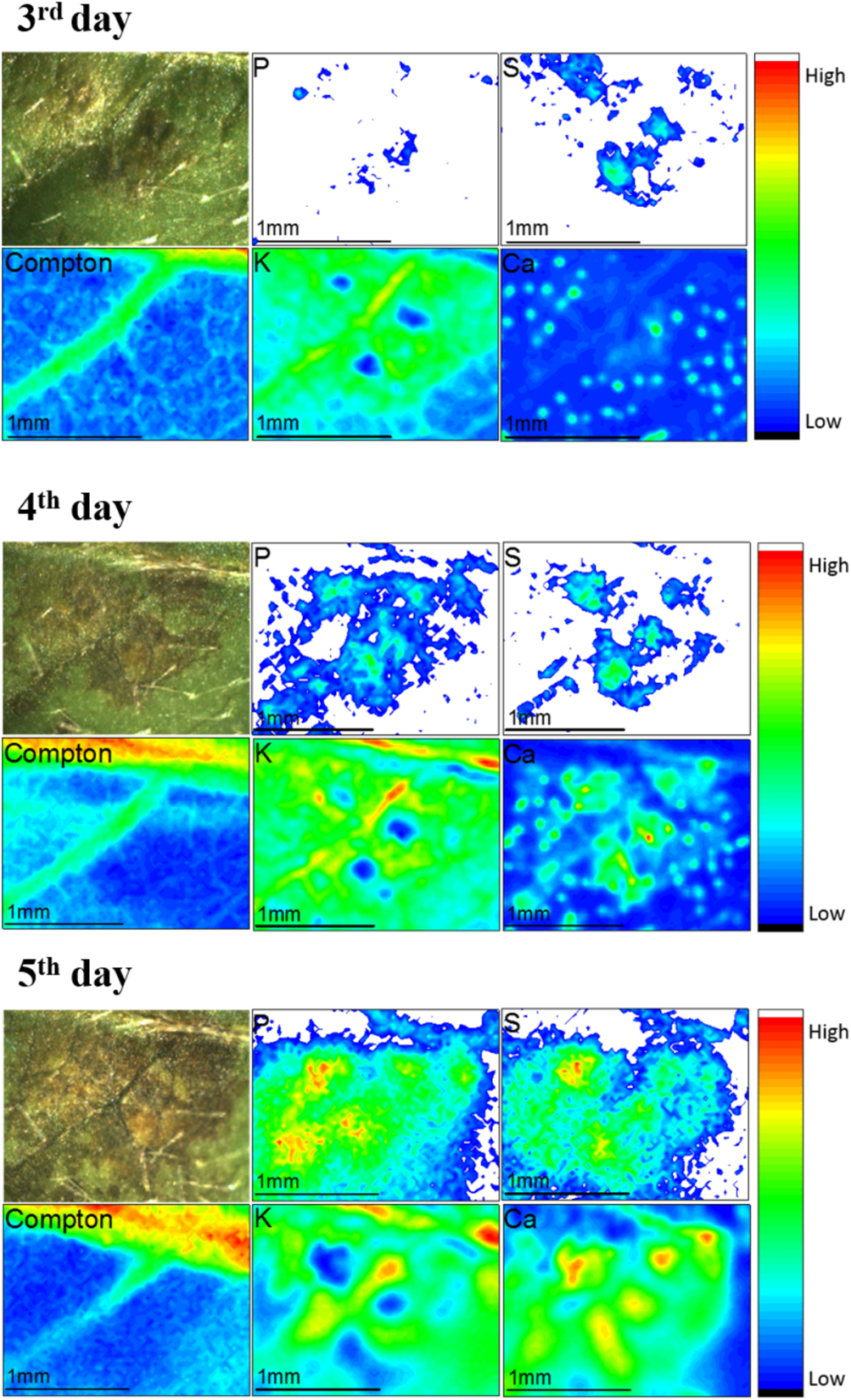
Figure 1: Micro XRF elemental mapping of the soybean leaf infected by anthracnose disease from the 3rd, 4th and 5th day after fungus Colletotrichum truncatum inoculation
Citation: Frontiers in Plant Science. 9, DOI: 10.3389/fpls.2018.01588.
---------------------------------------------------
Article by Chomphunuch Songsiriritthigul (Beamline Scientist, SLRI)
References
Bonto, A., Jearanaikoon, N., Sreenivasulu, N., Camacho, D. (2020). High uptake and inward diffusion of iron fortificant in ultrasonicated milled rice. LWT-Food Science and Technology. 128(5), 109459. DOI: 10.1016/j.lwt.2020.109459.
Mongkhonsin, B., Nakbanpote, W., Hokura, A., Nuengchamnong, N., Maneechai, S. (2016). Phenolic compounds responding to zinc and/or cadmium treatments in Gynura pseudochina (L.) DC. extracts and biomass. Plant Physiology and Biochemistry. 10, 549-560. DOI: 10.1016/j.plaphy.2016.10.027.
Nakhforoosh, A., Hallin, E., Karunakaran, C., Korbas, M., Stobbs, J., Kochian, L. (2024). Visualization and Quantitative Evaluation of Functional Structures of Soybean Root Nodules via Synchrotron X-ray Imaging. Plant Phenomics. 6, 0203. DOI: 10.34133/plantphenomics.0203.
Navarro, H., Marcó, L. M., Araneda, A. A., & Bennun, L. (2019). Spatial distribution of Si in Pinus Insigne (Pinus radiata) Wood using micro XRF by Synchrotron Radiation. Journal of Wood Chemistry and Technology, 39(3), 187–197. DOI: 10.1080/02773813.2018.1562473.
Rodrigues, E. S., Gomes, M. H. F., Duran, N. M., Cassanji, J. G. B., da Cruz, T. N. M., Sant’Anna Neto, A., Savassa, S. M., de Almeida, E., Carvalho, H. W. P. (2018). Laboratory Microprobe X-Ray Fluorescence in Plant Science: Emerging Applications and Case Studies. Frontiers in Plant Science. 9, DOI: 10.3389/fpls.2018.01588.
Vives, A. E. S., Silva, R. M. C., Medeiros, J. G. da S., Tomazello Filho, M., Barroso, R. C., Zucchi, O. L. A. D., & Moreira, S. (2005). Accumulation of elements in annual tree rings measured by synchrotron x‐ray fluorescence analysis. X-Ray Spectrometry, 34(5), 411–416. DOI: 10.1002/xrs.844.

111 SirinthonWitchothai Building, University Avenue, Muang District, Nakhon Ratchasima 30000, THAILAND
Tel.: +66 44 217 040 Fax: +66 44 217 047 Email: siampl@slri.or.th
Copyright © 2015 Synchrotron light research institute. All Rights Reserved.
|
|