Welding high vacuum parts is knowledge developed by the Synchrotron Light Research Institute, as these welded samples are essential equipment for accelerators and beamlines. Therefore, the features and quality of the samples must be inspected to evaluate the performance of the vacuum materials produced.
An example of welding vacuum materials at SLRI's "Instrument Fabrication Section” which we designed and developed, is the welding of austenitic stainless steel AISI304 samples with oxygen-free copper (OFHC copper) using the Gas Tungsten Arc Welding process. This process produces sample materials with the ability to transfer heat and are strong, making them suitable for using in various devices in accelerators and beamline systems, such as photon beam masks and photon beam slits.
These vacuum materials are fabricated by welding together different metals. Scientists brought the samples to beamline 7.2W: Macromolecular Crystallography (MX) for feature and quality checks, using hard X-ray synchrotron light from the Macromolecular Crystallography beamline, obtained from a 6.5 Tesla superconducting wavelength shifter. The light is focused with an X-ray polycapillary half lens, resulting in a focal point of 42 microns, sufficient for distinguishing each area of the welded part.
Scientists employed micro-X-ray fluorescence spectroscopy (XRF) and micro-X-ray absorption spectroscopy (XAS) technique to study the welded AISI304 stainless steel and copper, as these techniques are suitable for examining the type of element and its composition in small areas on the nonhomogeneous sample.
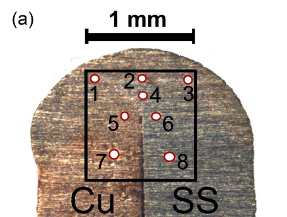
(Figure a) Sample welded between AISI304 stainless steel and copper with 8 analysis areas.
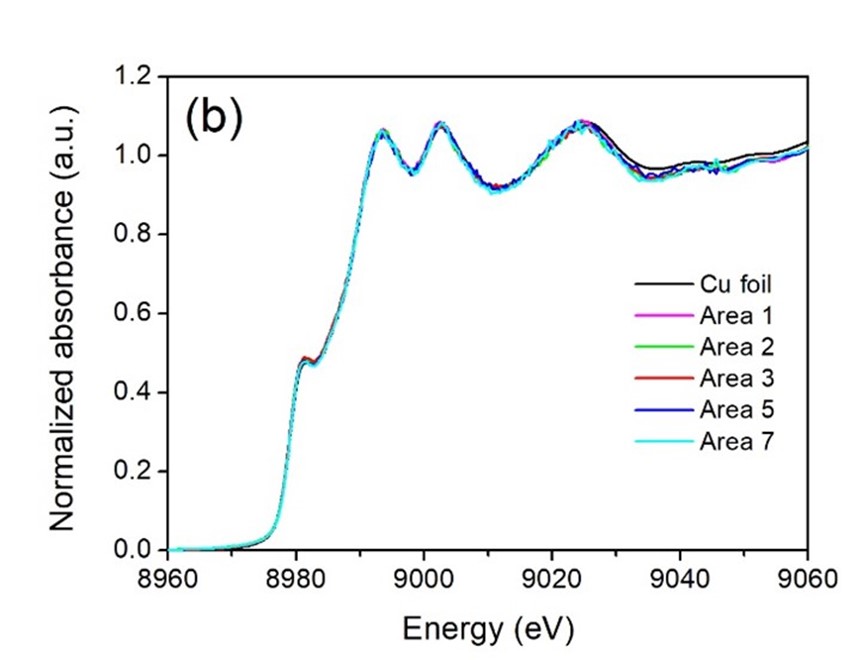
(Figure B) X-ray absorption spectra at the K-edge absorption region of various elements tested at Beamline 7.2, SLRI.
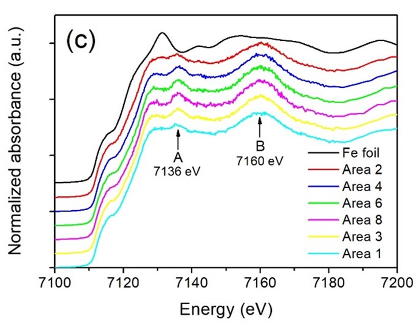
(Figure C) X-ray absorption spectra at the K-edge absorption region of various elements tested at Beamline 7.2, SLRI.

(Figure D) X-ray absorption spectra at the K-edge absorption region of various elements tested at Beamline 7.2, SLRI.
The examination of the sample from Figure A with synchrotron light at beamline 7.2W reveals the X-ray absorption spectrum at the K-edge absorption area of various elements, as depicted in Figures B, C, and D. The spectral characteristics of copper in various areas showed no difference (Figure B). However, the results exhibit a distinct pattern in the steel spectrum, as seen in Figure C, between the stainless-steel area (Area 8, SS side) and the welding joint area (Area 2), characterized by the presence of peaks A and B in the former.
The results indicate that Manganese, comprising 2 percent by weight, could replace iron atoms, exhibiting a structure resembling Face-Centered Cubic. This is evident in the stainless-steel area (Area 8, SS side) as shown in Figure D.
The sample examination is a result of continuous development and improvement of the beamline system, aimed at utilizing synchrotron light cost-effectively and efficiently. The high intensity X-ray focused to a small scale at the micrometer level is useful in distinguishing various features within small areas of a sample. Moreover, it is highly probable to be applied as a tool for analyzing more diverse types of samples in the future.
Article by: Chomphunuch Songsiriritthigul, Beamline Scientist
Complier: Corporate Communications Section
Translation: International Relations Officer

111 SirinthonWitchothai Building, University Avenue, Muang District, Nakhon Ratchasima 30000, THAILAND
Tel.: +66 44 217 040 Fax: +66 44 217 047 Email: siampl@slri.or.th
Copyright © 2015 Synchrotron light research institute. All Rights Reserved.
|
|