

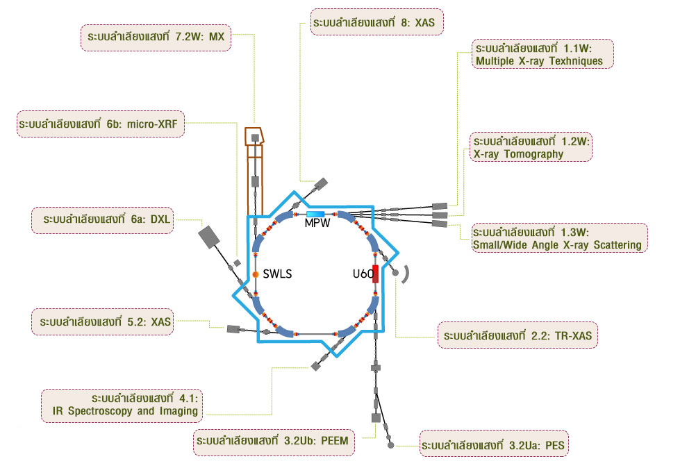




Time-resolved XAS Beamline Technology
The TRXAS beamline (BL2.2) employs an energy-dispersive monochromator (EDM) based on a bent Si(111) crystal, which horizontally focuses the polychromatic X-ray beam to a focal spot of approximately 1 mm at the sample position. This energy-dispersive configuration enables the entire X-ray Absorption Near Edge Structure (XANES) region to be collected simultaneously, without mechanical energy scanning.
ED-XAS (Energy-dispersive X-ray Absorption Spectroscopy)
The TRXAS beamline (BL2.2) was established and commissioned in 2012, utilizing the ED-XAS (Energy-Dispersive X-ray Absorption Spectroscopy) scheme. Key hardware, including the energy-dispersive monochromator (EDM) and a 2-theta arm, was donated by the University of Bonn, Germany.
- Design Strategy
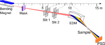
To compensate for the absence of an optical mirror, the beamline was designed with a very short physical length (approx. 9.7 m). This maximizes photon flux and provides a large acceptance angle of 8 mrad, which in turn allows for a broader X-ray energy range.
- Monochromator
Uses an Energy Dispersive Monochromator (EDM) with bent Si(111) and Si(220) crystals to focus a polychromatic beam onto the sample.
- Operating Range
Wide 2θ range (30° – 55°) supporting 4–12 keV, covering Ti K-edge to Pt L3-edge.
- Flexibility
The elliptical curvature of the crystal is precisely adjustable via a three-axis goniometer.
TRXAS (Time-resolved X-ray Absorption Spectroscopy)
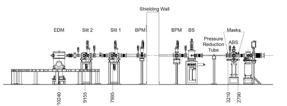
Schematic of the TRXAS beamline showing the main components, namely the heat absorber (ABS), front-end mask, differential-pressure tube, beam shutter (BS), retractable water-cooled Be foil, beam-position mornitor (BPM), two sets of vertical and horizontal slits and EDM. Distances from the source (in mm) are also shown.
Performance and Scanning Speed
The scanning speed performance of the endstation was evaluated by measuring the X-ray absorption of a 5 μm thick Ti foil. The Ti K-edge XANES spectra successfully resolved the characteristic absorption features of metallic titanium with a temporal resolution as fast as 25 ms, although with a relatively low signal-to-noise ratio.
Data quality improves significantly as the acquisition time increases to 200 ms or more. Based on the current synchrotron beam quality, the optimal recording speed for high-quality spectra typically ranges between 500 ms and 1 second.
Beamline Specification
| Parameter | Specification |
| X-ray Source | Bending Magnet (1.2 T) |
| Energy Range | 4.0 – 12.0 keV |
| Beam Size | 1 mm (H) x 2 mm (V) |
| Beam Divergence at Sample (mead)(FWHM) | |
| Monochromator | Bent Crystal Si(111) and Si(220) (Optional) |
| Min. Acquisition Time | 25 ms per spectrum |
| Detector | NMOS with Scintillator (1024 pixels) |
| Optical Bench | 2.8 m long |