Facility and Equipment Availability
Facility and Equipment Availability
Beamline 1.2W · X-ray Tomographic Microscopy (XTM)
Facility and Equipment Availability
Sample holders · Beamline Operation · Beamline Manual
Available sample holders & tools
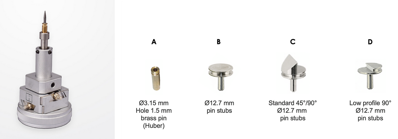
- Sample holder options at BL1.2W
- 45° Mount, Ø = 6 mm, aluminum
- 45° and 90° Mount, Ø = 6 mm, aluminum
- 90° Mount , Ø = 6 mm , aluminum
- Ø = 6 mm , aluminum
- Ø = 1.5 mm, brass
- Handling and mounting use container density less than sample density. Often used are small polystyrene tubes
- In order to mount the sample to sample holder use transparent glue, wax or 2-side tape
- User should make sure that the sample is stabilized to the sample holder other wise sample displacement during data collection will affect the CT reconstruction
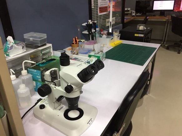
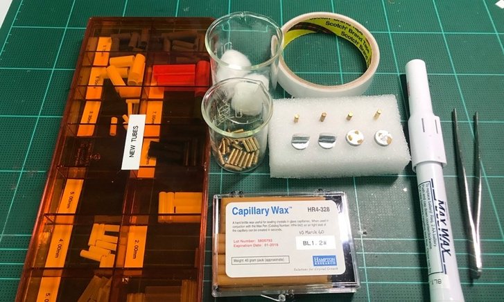
Other apparatus that may be needed in sample preparation e.g. heat gun, glue gun, and stereo microscope, are also provided at the Sample Station
Beamline Operation
Personal Safety Interlock (PSI) System
IMPORTANT: Before operating BL1.2W, users must acknowledge this local regulation:
- To protect the radiation from the experimental hutch, user must be trained for Beamline Hutch Search and Secure and Breaking Security
- Operation of the beamline must only be undertaken by authorized person
- User must wear a provided radiation-controlled badge at all times in the experimental hall
- User should not remain in the experimental hall during beam injection
- No food or drinks are allowed at the beamline station
- Users should clean up beamline on the last day of beamtime
PERSONNEL SAFETY SYSTEM (PSS)
PPS consist of electrical interlocks and mechanical barriers & locks that prevent personnel from entering beam enclosures and other areas in which the potential for high radiation exposure exists so that they are protected against prompt radiation. The interlocks also serve to shut off the radiation source if any of the interlocked barriers into beam enclosures are breached.
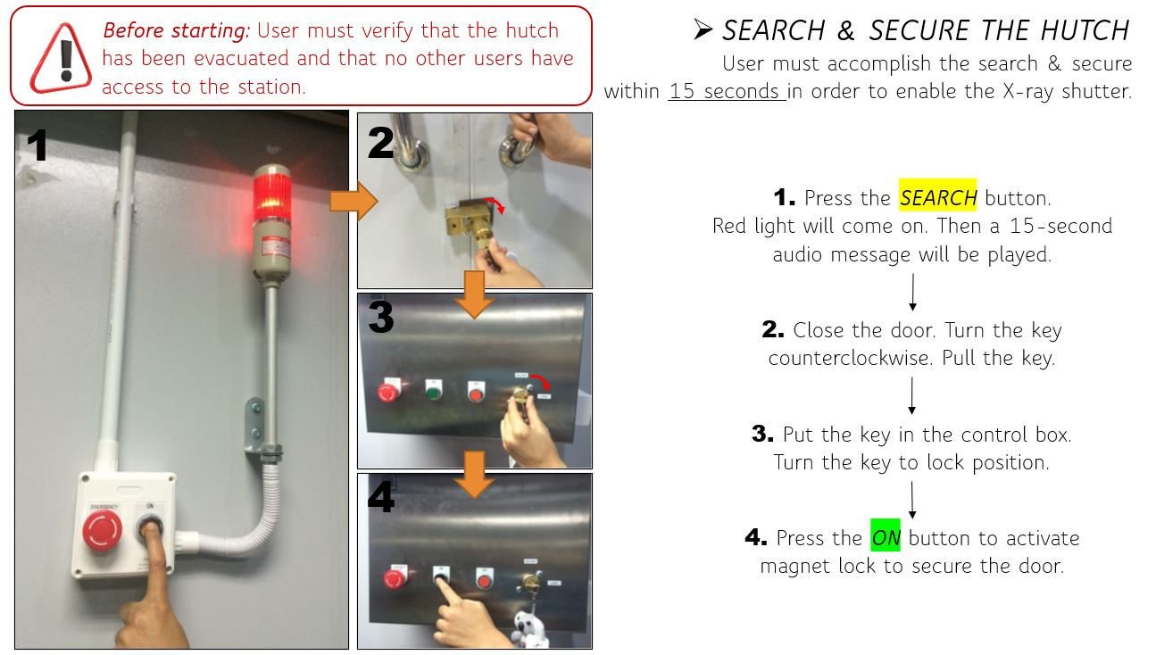
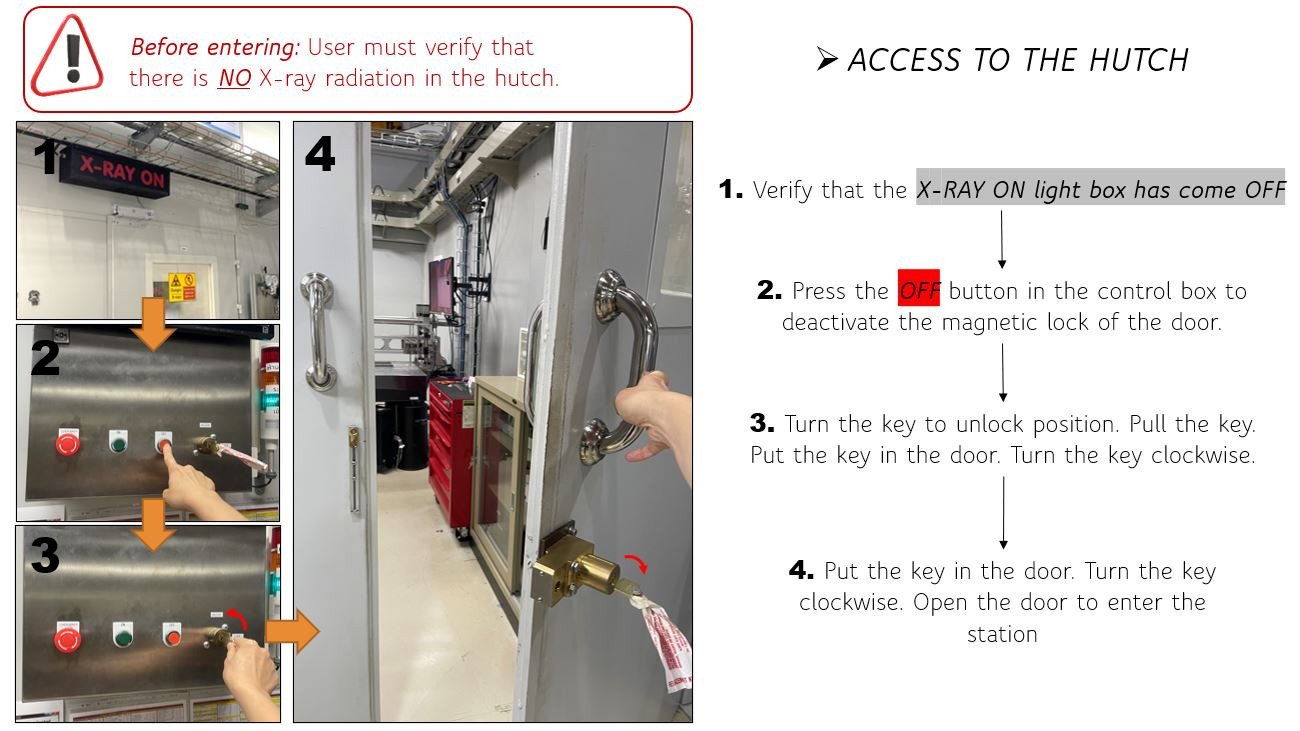
Search & secure the hutch
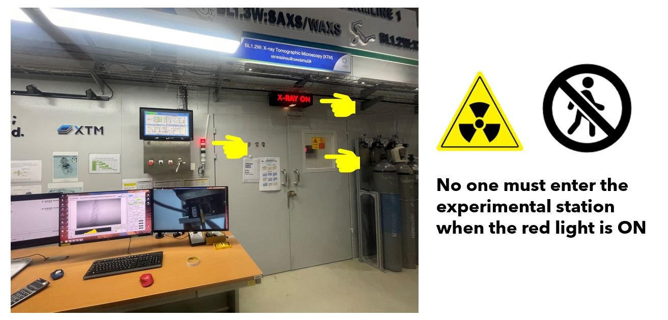
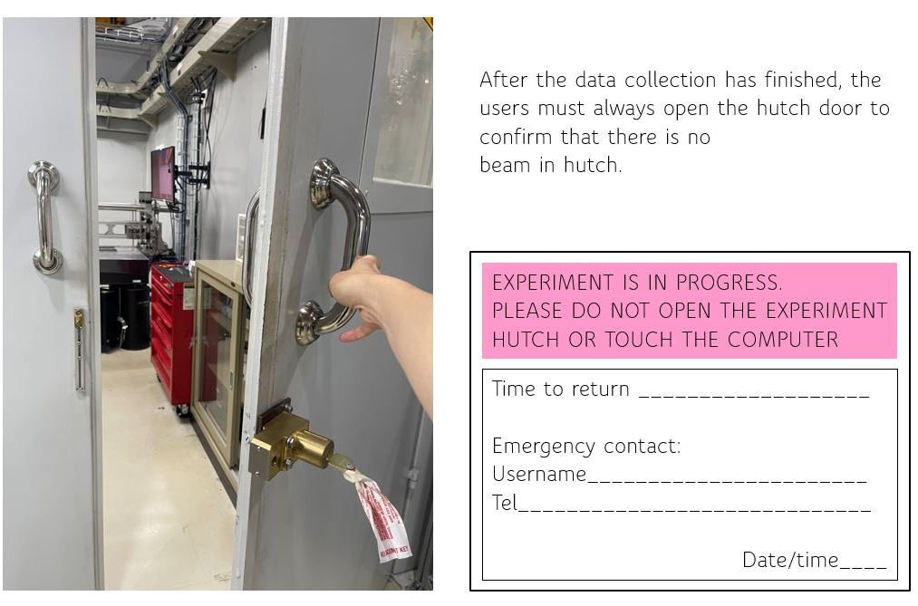
Before beam is allowed into an experimental enclosure, a search & secure procedure must be performed by the user to ensure that no individual is in the experimental station during beam transmission through the experimental beamline.
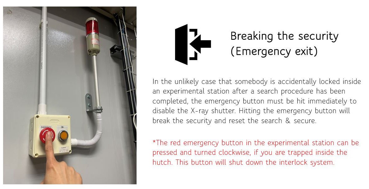
Breaking the security (Emergency exit)
In the unlikely case that somebody is accidentally locked inside an experimental station after a search procedure has been completed. Emergency exit procedure will break the security and reset the search & secure.
Beamline Manual
Sample Preparation
SRXTM at XTM beamline is based on microtomography geometry with an available x-ray energy range of 5 – 15 keV. To obtain the best imaging quality, the sample must be fixed or stabilized to make sure that there is no displacement during tomographic imaging. We highly recommend the samples be prepared in a cylindrical shape to minimize the imaging artifacts. For inorganic samples (composed of Ca, Cu, Se, Fe, Zn, etc.) or higly absorbe x-rays, the size should be less than 3 mm in diameter. However, for other samples that are composed of organic elements (C, H, O, N) or poorly absorb x-rays, the size can be up to 10 mm in diameter. SRXTM at XTM beamline is not suitable for larger samples, alloys, or titanium composites due to limited x-ray energy. Feasible samples are listed below unless stated otherwise.
SIZE of the sample
For an optimal absorption contrast, the x-ray images are aimed to obtain at 30% transmission (or 70% absorption). The absorption of X-rays by a sample depends on the quality of the X-ray beam, the elemental composition in the sample as well as the density and thickness of this sample. In general, the sample should be aligned within the FOV during rotation. This mean that the appropriate sample must be smaller than the FOV listed in the optical performance table. For example, to obtain the highest-resolution images with the pixel size of 0.72 micron, the diameter or thickness of the sample must not exceed 1.7 mm. Unless it contains mostly organic compounds and/or poorly absorbs x-rays, the sample can be larger than FOV.
In case of sample larger than FOV, the tomography dataset will be acquired from concatenated scans following the image stitching procedure. The experiment should be setup carefully to avoid misalignment that would affect the tomographic calculation. Also, the increased data volume should be taken into account.
For further analysis such as % porosity or pore distribution, we recommend that the sample should be >10-times larger than the selected feature to justify the analysis. For example, to determine the porosity, a bone sample with an average pore size of 100 microns should be prepared in 2 – 3 mm in diameter.
SHAPE of the sample
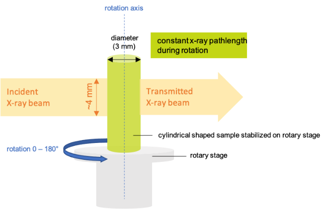
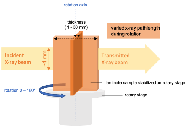
Cylindrical shaped sample is the most preferrable sample to x-ray tomography. According to the experimental setup, the x-ray beam is perpendicular to the rotation axis, the cylindrical shaped sample will give the constant pathlength (the width) during the sample rotation. This satisfies the back projection algorithm in tomographic reconstruction. In case of laminate sample, the pathlength will vary during rotation. Particularly when it turns the maximum width to the x-ray beam, it could give 0% transmission that disturbs the back projection algorithm. If it is possible, we suggest preparing the sample into a stick as illustrated.
List of feasible samples (size in thickness or diameter)
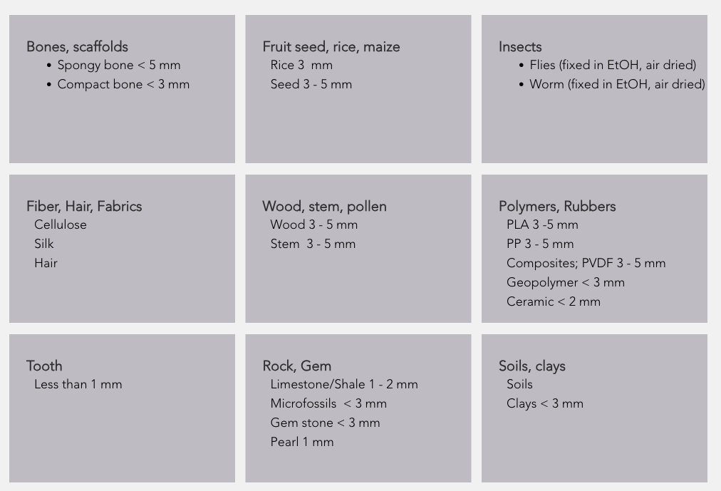
รายละเอียด sample holder ดูได้ที่หัวข้อ Available sample holders & tools ด้านบน
Instruction Manual
DATA PROCESSING
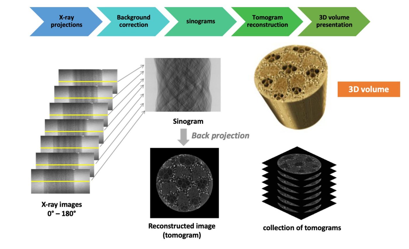
Reconstruction
Octopus Reconstruction Manual Octopus Recon user manual.pdf
Analysis
Octopus analysis manual.pdf
Visualization
Octopus visualazation manual.pdf
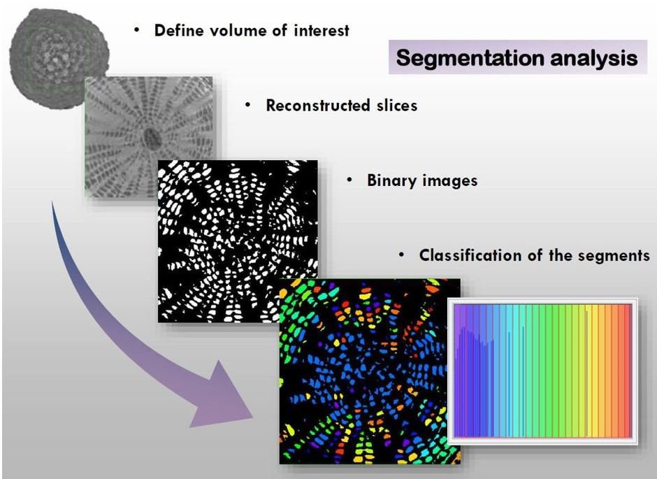
Quantitative analysis in tomography is based on binary images. At a given threshold, t generated by applying a threshold to the histogram of reconstructed slices. Binary images is resulting in binary image
Octopus Analysis Manual Octopus analysis user manual.pdf
Octopus Visualization Manual Octopus visualazation user manual.pdf
3D Volume
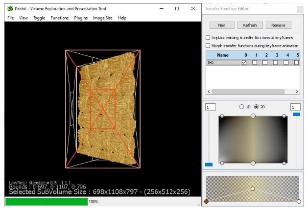
Drishti tutorials
The Basic of Drishti Drishti
3D Software Downlond https://github.com/nci/drishti/releases/tag/v2.6.5win