
Beamline Layout and Specification
Beamline Layout and Specification
Beamline Layout and Specification
Time-resolved XAS Beamline Technology
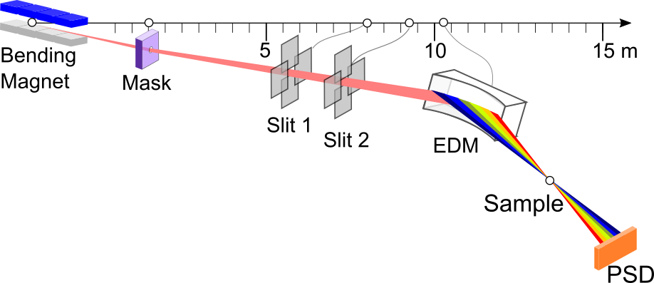
The TRXAS beamline (BL2.2) employs an energy-dispersive monochromator (EDM) based on a bent Si(111) crystal, which horizontally focuses the polychromatic X-ray beam to a focal spot of approximately 1 mm at the sample position. This energy-dispersive configuration enables the entire X-ray Absorption Near Edge Structure (XANES) region to be collected simultaneously, without mechanical energy scanning.
ED-XAS (Energy-dispersive X-ray Absorption Spectroscopy)
The TRXAS beamline (BL2.2) was established and commissioned in 2012, utilizing the ED-XAS (Energy-Dispersive X-ray Absorption Spectroscopy) scheme. Key hardware, including the energy-dispersive monochromator (EDM) and a 2-theta arm, was donated by the University of Bonn, Germany.
Design Strategy
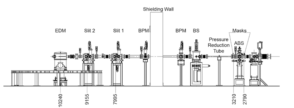
To compensate for the absence of an optical mirror, the beamline was designed with a very short physical length (approx. 9.7 m). This maximizes photon flux and provides a large acceptance angle of 8 mrad, which in turn allows for a broader X-ray energy range.
Monochromator
Uses an Energy Dispersive Monochromator (EDM) with bent Si(111) and Si(220) crystals to focus a polychromatic beam onto the sample.
Operating Range
Wide 2θ range (30° – 55°) supporting 4–12 keV, covering Ti K-edge to Pt L3-edge.
Flexibility
The elliptical curvature of the crystal is precisely adjustable via a three-axis goniometer.
TRXAS (Time-resolved X-ray Absorption Spectroscopy)
Performance and Scanning Speed
The scanning speed performance of the endstation was evaluated by measuring the X-ray absorption of a 5 μm thick Ti foil. The Ti K-edge XANES spectra successfully resolved the characteristic absorption features of metallic titanium with a temporal resolution as fast as 25 ms, although with a relatively low signal-to-noise ratio.
Data quality improves significantly as the acquisition time increases to 200 ms or more. Based on the current synchrotron beam quality, the optimal recording speed for high-quality spectra typically ranges between 500 ms and 1 second.
Beamline Specification
| X-ray Source | Bending Magnet (1.2 T) |
|---|---|
| Energy Range | 4.0 – 12.0 keV |
| Beam Size | 1 mm (H) × 2 mm (V) |
| Monochromator | Bent Crystal Si(111) and Si(220) (Optional) |
| Min. Acquisition Time | 25 ms per spectrum |
| Detector | NMOS with Scintillator (1024 pixels) |
| Optical Bench | 2.8 m long |