Facility and Equipment Availability
Facility and Equipment Availability
Beamline 3.2Ua/b · Photoemission Spectroscopy (PES) & PEEM
Facility and Equipment Availability
Equipment at each end station & Beamline Manual
ARPES End Station
| Equipment or Component | Details |
|---|---|
| Scienta R4000 hemispherical electron energy analyzer with +/- 15° angle multiplexing lens. | Analyzer entrance slit vertical, along main rotation axis of manipulator for Fermi surface maps. |
| Six-degree-of-freedom sample cryogenic manipulator (Gold sheet is installed on the manipulator) from Fermi Instruments | 5-axis X, Y, Z, R1 (polar), and R3 (tilt) with control motor and R2 (azimuth) with manual screw. XYZ and angular axis accuracy is 0.01 mm and 0.1 deg.
|
| Variable temperature 8 – 300 K | Take 8 hours from room temperature to 8 K. |
| Sample check and orientation by LEED | Maximum at 1 keV electron energy |
| Evaporation deposition on sample surface | Two slots of alkali metal evaporated source (K, Na, Li). Requirements need to be discussed with beamline scientists. |
| Ar ion sputtering gun | Maximum energy is 3 keV, and working pressure is ~10⁻⁶ mbar at preparation chamber |
| Gases flow into the preparation chamber | Ar, N₂, O₂, etc., gas flowing at the preparation chamber requirements need to be discussed with beamline scientists. |
| The MBE chamber is connected for annealing a sample or material deposition. | The maximum temperature of the heater is 700°C, with a pressure base of 10⁻⁹ mbar. Material deposition needs to be discussed with beamline scientists. |
| Load-lock with 7 slots of sample holders | Takes 1.5 hours to pump down for preparing pressure to transfer a sample into the main chamber without breaking UHV |
| Electrical connection with manipulator | Available to apply a voltage bias to a sample or measure current on a sample. |
| Fermi BL1200s Multi-Gas Ultra-High Flux VUV source system | An additional fixed VUV source component system with a He-discharged gas source and a monochromator generates photon energy of 21.22 eV |
XPS End Station
| Equipment or Component | Details |
|---|---|
| Hemispherical Electron Energy Analyzer: Thermo VG Scientific Alpha110 | Total minimum energy resolution at 200 meV and RT, and typical XPS Au4f7/2 FWHM is about 0.8 eV at the pass energy of 10 and photon energy of 400 eV |
| 4-axis manipulator | X, Y, Z, and R1 (polar) 0-180 deg |
| Ar ion sputtering gun | Maximum energy is 3 keV, and working pressure is ~10⁻⁶ mbar in the main chamber |
| Electron gun | The energy up to 5 keV |
| Load-lock with 2 slots of sample holder plate | Takes at least 1.5 hours (depending on the sample type) to pump down for preparing pressure to transfer a sample into the main chamber without breaking UHV |
| Total Fluorescence Yield detector | Hamamatsu photonics multi-channel plate |
| Electrical connection with manipulator | Available to apply a voltage bias to a sample or measure current on a sample. |
| Flux monitor | Gold mesh and photodiode |
| Large sample holder plate | 28 mm x 94 mm sample plate, able to load several samples in the main chamber (numbers of samples depend on the sample size). |
PEEM End Station
| Equipment or Component | Details |
|---|---|
| Hemispherical electron energy analyzer with Micro-channel plate, Sensicam QE CCD camera | Model: Elmitec PEEM III |
| Electron gun low-energy electron microscopy (LaB6 thermionic emission) | LEED pattern, LEED-IV, EELS incidence electron energy from -10 to 500 V |
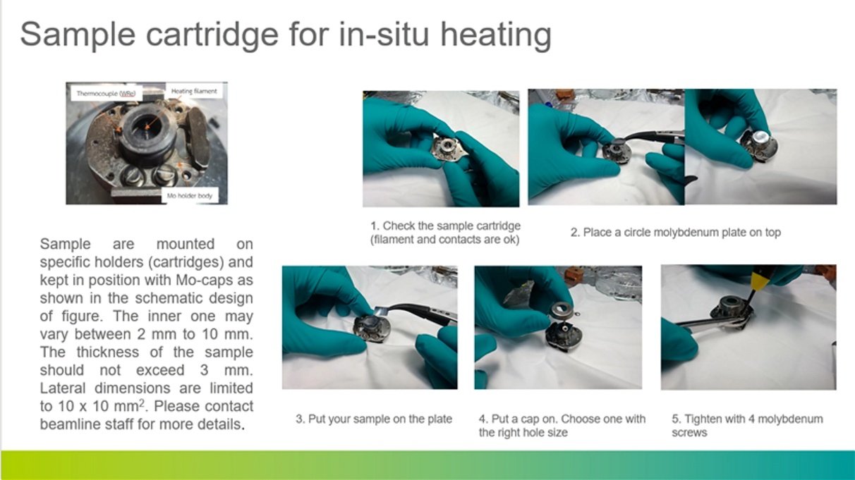
| 3-axis manipulator with heater | The sample can be heated by thermal filament up to 600°C |
| Electron bombardment heating | The sample temperature can be more than 1200°C at a maximum current |
| IR pyrometer | It detects IR radiation from the sample and converts it to temperature in real time using the sample’s emissivity. |
| Ar ion sputtering gun | Maximum energy is 3 keV, and working pressure is ~10⁻⁶ mbar in the preparation chamber. |
| E-beam evaporation cell | Requirements need to be discussed with beamline scientists. |
| Gases flow into the preparation and main chamber | Ar, N₂, O₂, etc., gas flowing requirements need to be discussed with beamline scientists. |
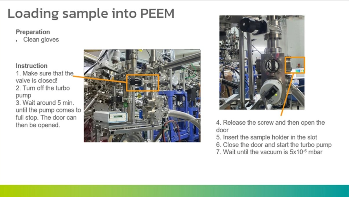
| Load-lock with 2 slots of sample holder | Takes at least 1.5 hours (depending on the sample type) to pump down for preparing pressure to transfer a sample into the main chamber without breaking UHV |
Support Facilities
| Equipment or Component | Details |
|---|---|
| Digital hotplate | Temperature range: 5-550°C, ± 2.0°C |
| Low-to-Mid Range Magnification Digital Microscope LEICA DMS1000 | The optics ensures magnification of up to 300x. The built-in HDMI microscope camera provides full high definition live images of up to 30 fps and a resolution of 5M pixels. |
| Ultrasonic cleaner | – |
| Silver epoxy, high temperature Carbon paste | – |
| Cu, Al, Ni plates | For powder sample substrate of powder specimen |
| Carbon and Copper tape | – |
| Ceramic and Al wire top-post | For cleavable samples |
| Sample holders | – |
| Desiccator Cabinet | – |
| Spot welding tool | – |
Beamline Manual
คลิกหัวข้อเพื่อเปิดอ่าน
BL3.2Ua (PES) has two photoemission experimental stations using the synchrotron radiation. PES is for angle-resolved photoemission, and XPS is for angle-integrated photoemission. Sample requirements are different for each station. Basically, XPS is opened for those who want to measure spectra on many samples, including fine powders and large-sized materials, at room temperature. PES has a limited capacity for samples, but the in-situ heating and surface metal deposition are available.
1. ARPES Station
The sample is mounted on the holder (see a drawing below) by silver adhesive paste, and loaded into the 6-axis movable and cryo-cooled manipulator (Fermi instruments). A sample holder covered by a thermal radiation shield can be cooled down to 8 K from room temperature overnight. The Ar ion gun (3 keV) is also available for in-situ surface treatment.
1.1 Sample requirements
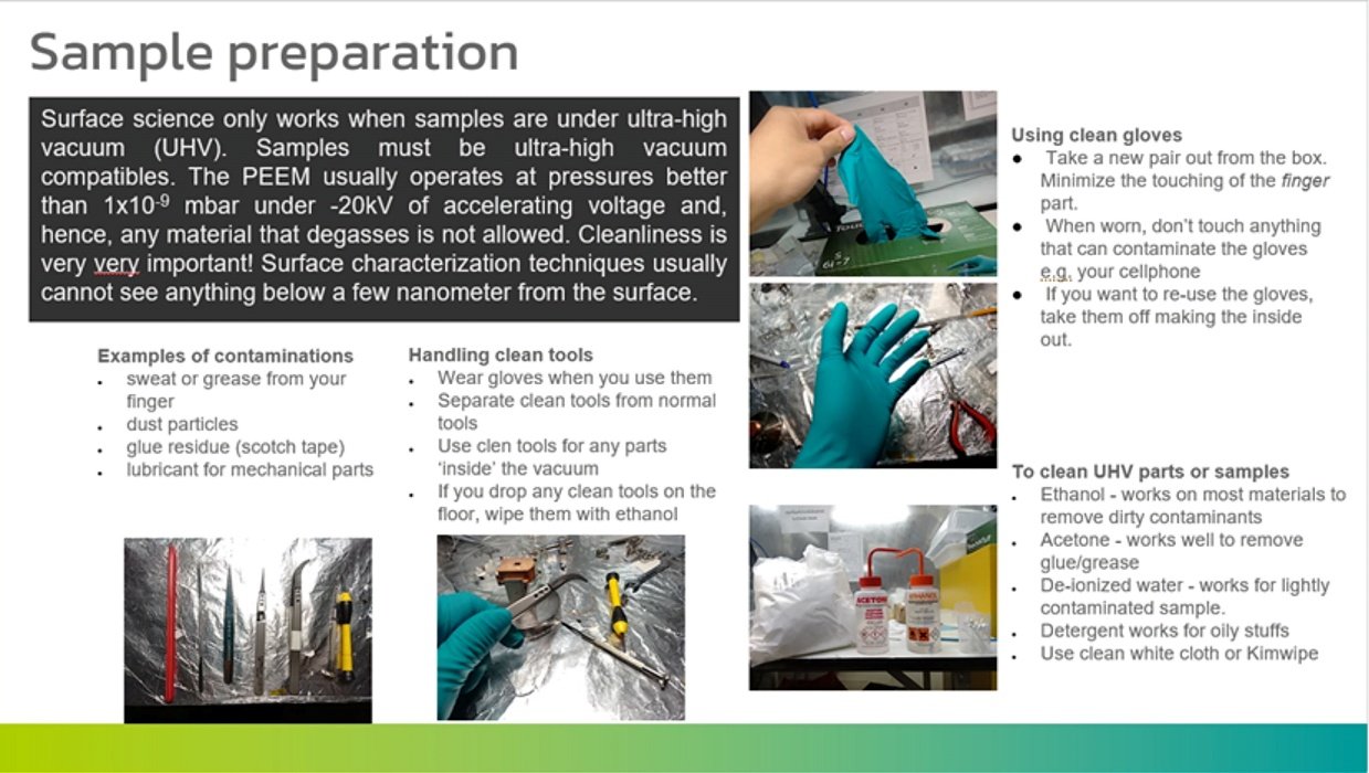
- The sample must be compatible with ultra-high-vacuum conditions
- Cleavable material (usually a layered crystal structure) or single-crystalline material
- The sample surface must be sufficiently conductive
- The size of the sample should be more than 2-5 mm in diameter
1.2 Sample preparation
Samples have to be prepared in situ under ultra-high vacuum to produce clean, atomically controlled surfaces. In the vacuum system, the following techniques will be available:
- Cleavage of layered materials by knocking off a post-silver glued to the top surface.
- Ar ion gun sputtering (3 keV @ max energy)
- Annealing at separated MBE chamber
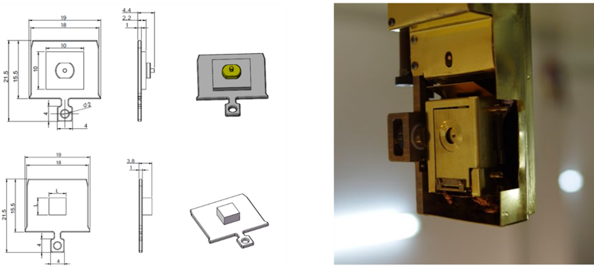
Sample holder dimension and manipulator
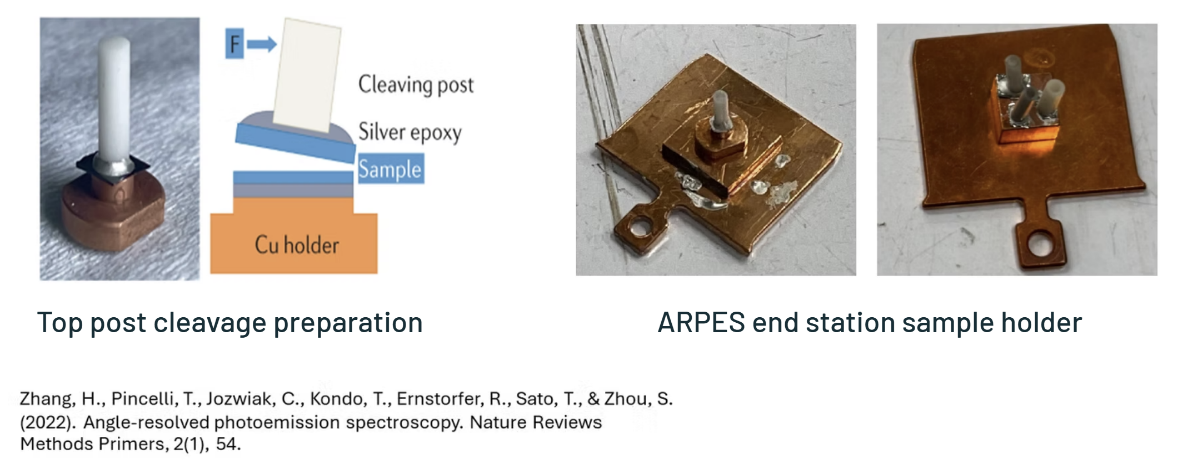
Sample preparation
1.3 Data analysis
Data analysis software “Igor Pro” is available on PCs at the ARPES station. This is a commercial software licensed only at the station. Simple data analysis can be performed using Microsoft Excel 2007 (contact the beamline staff for the Microsoft Excel macro). Users are recommended to purchase the software either “Igor Pro” or “Origin” for further analysis in your office.
2. XPS Station
The sample is mounted on the stainless plate of 28 mm x 94 mm x 2 mm (thickness), sliding into the home-developed sample transfer mechanism based on the manipulator: HPT x-y, z translator. Sample surfaces should be conductive by either silver adhesive (12686-15: Silver Conductive Adhesive 503) or epoxy (H21D). Powder samples are attached on carbon adhesive tape (6 mm x 20 mm). There are 2 slots for sample holders in the load-lock chamber. Samples loading duration before transfer to the measurement chamber is about 40 min., but it depends on your sample condition. For example, if the samples are powder shape, it takes at least one hour to load the samples into the analysis. The sample surface can be sputtered by a 3 keV Ar ion at most in-situ prior to XPS. No in-situ sample heater is available. A sample drain current can also be measured in NEXAFS mode.
2.1 Sample preparation and requirements
- The sample must be compatible with ultra-high-vacuum conditions
- The sample surface must be sufficiently conductive
- The size of the sample should be more than 3 mm in diameter and less than 5 mm thick.
- The analytical area is about 1 mm x 1 mm.
- The proper size is about 5 mm x 5 mm, within 1 mm thick.
- A control or standard sample should be prepared by users for reference.
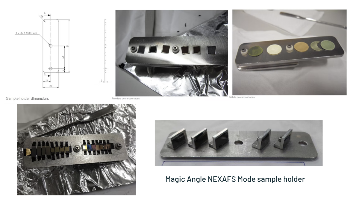
2.2 Data analysis
Data analysis software “Avantage” is available on PCs at the XPS station. This is a commercial software licensed only at the station. Simple data analysis can be performed using Microsoft Excel 2007 (contact the beamline staff for the Microsoft Excel macro). Users are recommended to purchase the software either “Igor Pro” or “Origin” for further analysis in your office.
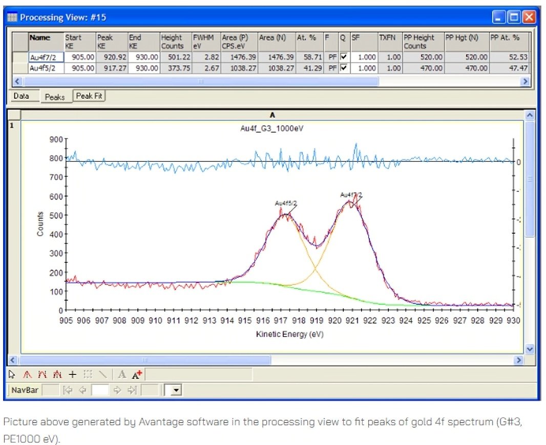
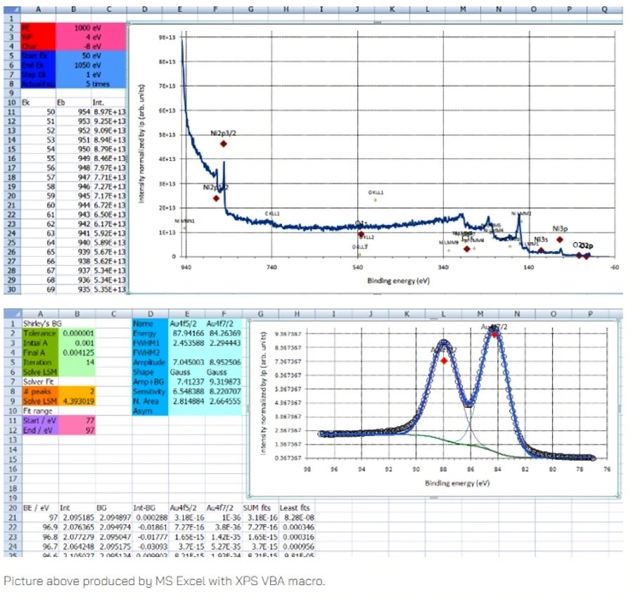
Contact the beamline staff for the Microsoft Excel macro
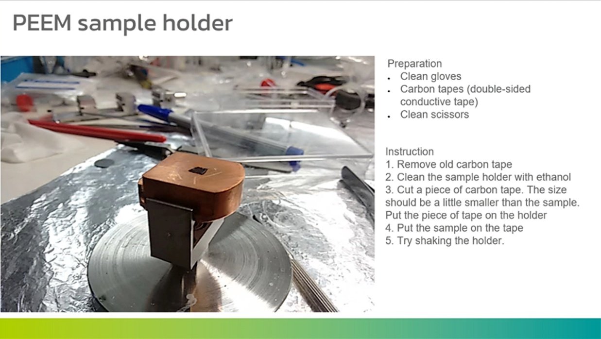
3. PEEM Station