Beamline8: XAS/XFI
X-ray Absorption Spectroscopy and X-ray Fluorescence
What is X-ray Absorption Spectroscopy (XAS)?
X-ray Absorption Spectroscopy (XAS) is an advanced spectroscopic technique that uses tunable X-rays typically generated from synchrotron radiation—to probe the local environment of specific atoms within a material. XAS provides insight into:
- Oxidation states
- Local atomic structure
- Chemical bonding and symmetry
- Electronic states of the absorbing atom
Why Use XAS
XAS is a powerful technique because it:
- Requires minimal sample preparation and is applicable to solids, liquids, gases, and solutions
- Offers element-specific analysis
- Provides atomic-scale structural information
- Can analyze highly diluted samples (down to ppm levels) and multiphase systems
- Allows in situ and operando measurements of materials under working conditions
What is X-ray Fluorescence Imaging (XFI)?
XFI is an analytical technique that uses X-ray excitation to map the spatial distribution of elements within a sample.
When a sample is irradiated by an X-ray beam, atoms in the sample emit characteristic fluorescence X-rays.
These emitted X-rays are detected and analyzed to determine the elemental composition at different positions,
creating an image that shows how elements are distributed across the sample.
Key features:
- Non-destructive: Suitable for delicate or historical samples.
- Element-specific: Each element emits a unique fluorescence lines.
- Spatially resolved: Provides micrometer-scale resolution of elemental distribution.
Two main approaches:
- Micro-beam XFI (µXFI): Uses a focused X-ray beam and scans the sample pixel by pixel.
- Full-field XFI (FXI): Uses a large, unfocused X-ray beam and a position-sensitive detector (e.g., pnCCD), capturing the entire field simultaneously.
XAS at BL8 – the Synchrotron Light Research Institute (SLRI)
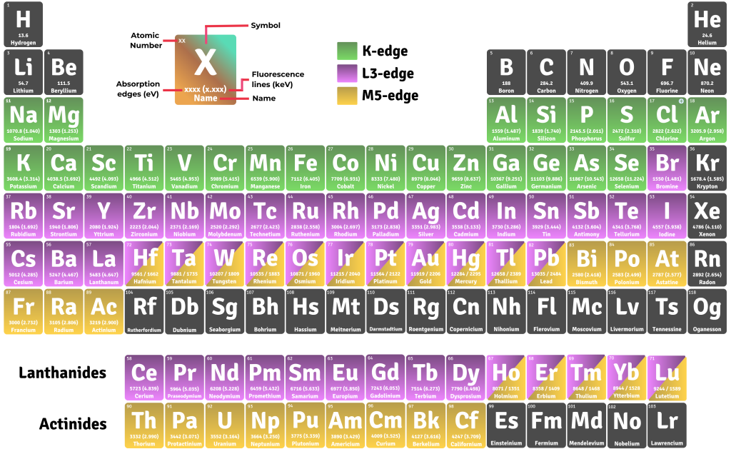
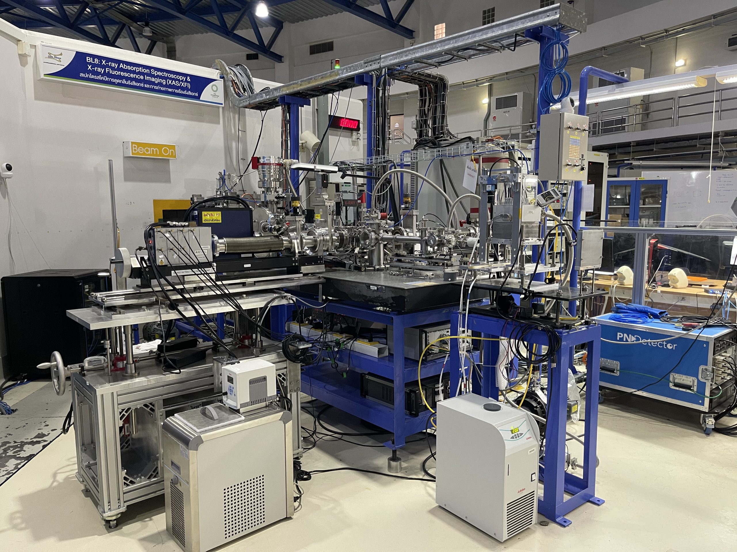
Beamline BL8 (XAS) at SLRI is a dedicated facility for X-ray Absorption Spectroscopy, supporting both XANES and EXAFS measurements. It operates in an energy range of 1–13 keV, suitable for studying the K-edges of elements from sodium to lead, as well as L- and M-edges of heavier elements.
- Oxidation states
- Local atomic structure
- Chemical bonding and symmetry
- Electronic states of the absorbing atom
Key Advantages of BL8 Compared to Other XAS Beamlines
Based on performance evaluations, BL8 offers:
- High photon flux: 10 8 – 10 11 photons/s/100 mA
- Excellent energy resolution: 3 × 10⁻⁴ – 4 × 10⁻⁴
- High energy stability: drift of approximately 0.07 eV
- Large beam size: up to 14 mm × 2 mm, ideal for bulk samples
Additional strengths include:
- Broad coverage of multiple absorption edges
- Multiple detection modes (Transmission and Fluorescence)
- High sensitivity for dilute systems, especially for P, S, and Cl
- High stability and precision, suitable for both fundamental and applied research in materials science, biology, environmental studies and archaeology
XFI at BL8 – Synchrotron Light Research Institute (SLRI)
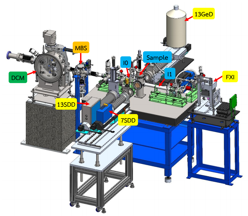
Full-field XRF Imaging (FXI)
- Uses a large, unfocused X-ray beam and detects the fluorescence signals with a pnCCD array detector, functioning like an X-ray camera.
- Captures the entire field simultaneously (simultaneous measurement), making it more time-efficient compared to µXFI.
- At BL8 SLRI, the X-ray beam size is 13 mm × 2 mm, tilted to achieve a footprint of approximately 13 mm × 15 mm.
Applications and Research Examples
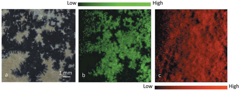
- Geology: Mapping Mn distribution in dendritic limestone.
- Archaeology: Investigating Sn and Cu segregation in ancient bronze.
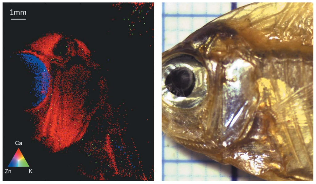
- Biology/Food Science: Analyzing Zn in fisheyes and Ca in fish bones.